

Time-resolved universal temperature measurements using NaYF4:Er3+,Yb3+ upconverting nanoparticles in an electrospray jet
- PMID: 30546988
- PMCID: PMC6278772
- DOI: 10.3762/bjnano.9.270
Time-resolved universal temperature measurements using NaYF4:Er3+,Yb3+ upconverting nanoparticles in an electrospray jet
Abstract
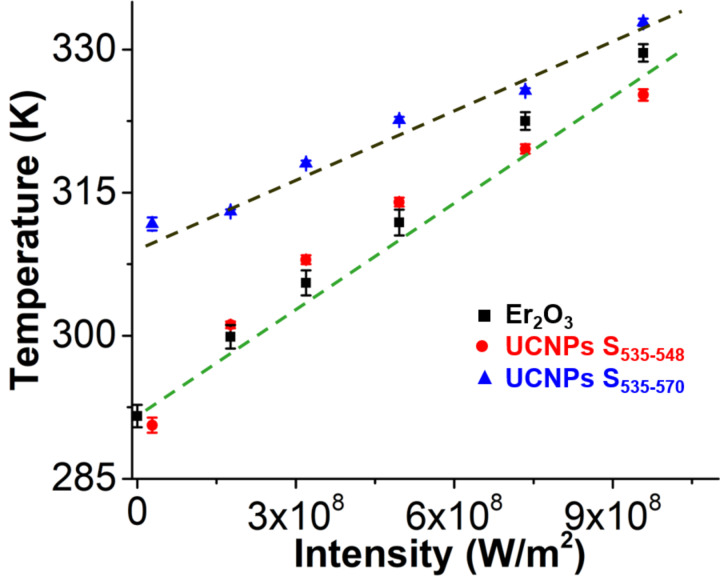
Hexagonal upconverting nanoparticles (UCNPs) of NaYF4:Er3+,Yb3+ (ca. 300 nm) have been widely used to measure the temperature at the nanoscale using luminescence ratio thermometry. However, several factors limit their applications. For example, changes in the peak shape, mainly is the S-band emission, hinders their ability to be used as a universal temperature sensor. Herein, we introduce a universal calibration protocol for NaYF4:Er3+,Yb3+ upconverting nanoparticles that is robust to environmental changes and gives a precise temperature measurement. We used this new procedure to calculate the temperature profile inside a Taylor cone generated with an electrospray jet. Inside the Taylor cone the fluid velocity increases toward the tip of the cone. A constant acquisition length leads to a decrease in excitation and acquisition time. This decrease in excitation time causes a peak shape change that corrupts the temperature measurement if the entire peak shape is integrated in the calibration. Our universal calibration circumvents this problem and can be used for time-resolved applications. The temperature at the end of the Taylor cone increases due to the creation of a whispering gallery mode cavity with 980 nm excitation. We use time-resolved energy balance equations to support our optical temperature measurements inside the Taylor cone. We believe that the findings of this paper provide a foundation for time-resolved temperature measurements using NaYF4:Er3+,Yb3+ upconverting nanoparticles and can be used to understand temperature-dependent reactions such as protein unfolding inside microjet/microdroplets and microfluidic systems.
Keywords: electrospray; microjet; nanothermometry; temperature measurement; time-resolved measurement; upconverting nanoparticles.
Figures


Similar articles
-
Monodisperse Core-Shell NaYF4:Yb3+/Er3+@NaYF4:Nd3+-PEG-GGGRGDSGGGY-NH2 Nanoparticles Excitable at 808 and 980 nm: Design, Surface Engineering, and Application in Life Sciences.Front Chem. 2020 Jun 12;8:497. doi: 10.3389/fchem.2020.00497. eCollection 2020. Front Chem. 2020. PMID: 32596210 Free PMC article.
-
Dual functional NaYF4:Yb3+, Er3+@NaYF4:Yb3+, Nd3+ core-shell nanoparticles for cell temperature sensing and imaging.Nanotechnology. 2018 Mar 2;29(9):094001. doi: 10.1088/1361-6528/aaa44a. Nanotechnology. 2018. PMID: 29283363
-
NaYF4:Er3+,Yb3+/SiO2 Core/Shell Upconverting Nanocrystals for Luminescence Thermometry up to 900 K.J Phys Chem C Nanomater Interfaces. 2017 Feb 16;121(6):3503-3510. doi: 10.1021/acs.jpcc.6b10279. Epub 2017 Jan 20. J Phys Chem C Nanomater Interfaces. 2017. PMID: 28303168 Free PMC article.
-
Turn-on detection of a cancer marker based on near-infrared luminescence energy transfer from NaYF4:Yb,Tm/NaGdF4 core-shell upconverting nanoparticles to gold nanorods.Langmuir. 2014 Nov 4;30(43):13085-91. doi: 10.1021/la502753e. Epub 2014 Oct 22. Langmuir. 2014. PMID: 25296290
-
Upconverting nanoparticles for nanoscale thermometry.Angew Chem Int Ed Engl. 2011 May 9;50(20):4546-51. doi: 10.1002/anie.201006835. Epub 2011 Apr 14. Angew Chem Int Ed Engl. 2011. PMID: 21495125 Review.
Cited by
-
Stabilization of Mixed-Halide Lead Perovskites Under Light by Photothermal Effects.J Energy Chem. 2021 Dec;63:8-11. doi: 10.1016/j.jechem.2021.08.046. Epub 2021 Aug 28. J Energy Chem. 2021. PMID: 35450060 Free PMC article.
References
LinkOut - more resources
Full Text Sources