

Case Reports
doi: 10.1016/j.ctro.2018.11.004.
eCollection 2019 Feb.
Radiation-induced vascular malformations in the brain, mimicking tumor in MRI-based treatment response assessment maps (TRAMs)
Affiliations
- PMID: 30547098
- PMCID: PMC6282630
- DOI: 10.1016/j.ctro.2018.11.004
Item in Clipboard
Case Reports
Radiation-induced vascular malformations in the brain, mimicking tumor in MRI-based treatment response assessment maps (TRAMs)
Clin Transl Radiat Oncol.
.
Abstract
•Of 310 brain tumors patients recruited, histology of 99 lesions was available.•Of those, 5 were histologically confirmed as radiation-induced malformations.•TRAMs cannot differentiate active tumor from vascular malformation.
Keywords: Brain tumors; MRI; RICM; RIT; Radiation; TRAMs; Vascular malformations.
Figures
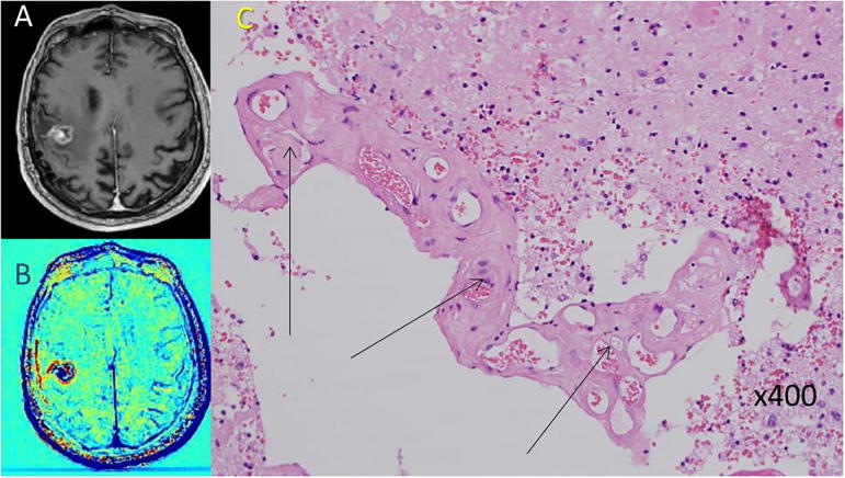
An example of radiation-induced vascular malformation in a patient with GBM post treatment (patient #2). A, B: Pre-surgical contrast-enhanced T1-weighted MRI and the calculated TRAMs depicting a small blue mass within a surrounding thin blue rim. C: H&E stained paraffin section showing radiation-induced cavernoma-like vascular malformation (arrows). (For interpretation of the references to colour in this figure legend, the reader is referred to the web version of this article.)
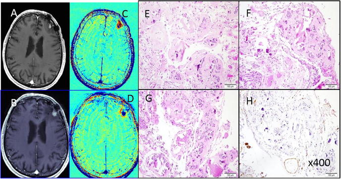
An example of radiation-induced vascular malformation in a patient with a non-small cell lung cancer brain metastasis post treatment (patient #3). A, C: Pre-surgical contrast-enhanced T1-weighted MRI and the calculated TRAMs depicting a small blue mass on the border of the previous surgery site 29 months post SRS. B, D: Pre-surgical contrast-enhanced T1-weighted MRI and the calculated TRAMs depicting a small blue mass re-growing on the border of the previous surgery site 23 months post removal of the previous lesion and FSR. E–H H&E stained paraffin sections showing cavernoma-like vascular malformations, with back to back vascular channels, marked hyalinization and calcification. H: Immuno-histochemical staining for smooth muscle alpha-actin (SMA) showing only patchy immuno-reactivity in vessel walls, with near-absence in areas. (For interpretation of the references to colour in this figure legend, the reader is referred to the web version of this article.)
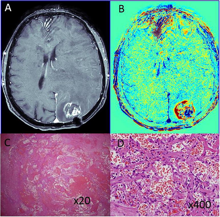
An example of radiation-induced vascular malformation in a patient with GBM post treatment (patient #4). A, B: Pre-surgical contrast-enhanced T1-weighted MRI and the calculated TRAMs depicting a small blue mass within a surrounding thin blue rim. C, D: H&E stained paraffin section showing radiation-induced cavernoma-like vascular malformation. (For interpretation of the references to colour in this figure legend, the reader is referred to the web version of this article.)
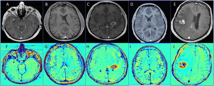
MRI of the patient 5–9 (patients with treated AVM). A–E: T1 of the five patients. F–J: TRAMs of the five patients.
Similar articles
-
Delayed contrast extravasation MRI: a new paradigm in neuro-oncology.Neuro Oncol. 2015 Mar;17(3):457-65. doi: 10.1093/neuonc/nou230. Epub 2014 Nov 30. Neuro Oncol. 2015. PMID: 25452395 Free PMC article.
-
Evaluating the diagnostic ability of treatment response assessment maps (TRAMs)/contrast clearance analysis (CCA) in predicting the presence of active brain tumors.Neuroradiol J. 2025 Feb 26:19714009251324305. doi: 10.1177/19714009251324305. Online ahead of print. Neuroradiol J. 2025. PMID: 40010303 Free PMC article.
-
Characterization of radiation-induced cavernous malformations and comparison with a nonradiation cavernous malformation cohort.J Neurosurg. 2015 May;122(5):1214-22. doi: 10.3171/2015.1.JNS141452. Epub 2015 Feb 20. J Neurosurg. 2015. PMID: 25699412
-
Intracranial extra-axial cavernous (HEM) angiomas: tumors or vascular malformations?J Neuroradiol. 2002 Jun;29(2):91-104. J Neuroradiol. 2002. PMID: 12297731 Review.
-
[Classification of superficial vascular anomalies].Presse Med. 2010 Apr;39(4):457-64. doi: 10.1016/j.lpm.2009.07.029. Epub 2010 Mar 4. Presse Med. 2010. PMID: 20206462 Review. French.
Cited by
-
Delayed Contrast-enhanced MRI Distinguishes Tumor from Radiation Treatment Effect.Radiol Imaging Cancer. 2025 May;7(3):e240388. doi: 10.1148/rycan.240388. Radiol Imaging Cancer. 2025. PMID: 40214516 Free PMC article. No abstract available.
-
Conventional and Advanced Imaging Techniques in Post-treatment Glioma Imaging.Front Radiol. 2022 Jun 28;2:883293. doi: 10.3389/fradi.2022.883293. eCollection 2022. Front Radiol. 2022. PMID: 37492665 Free PMC article. Review.
-
MRI Treatment Response Assessment Maps (TRAMs) for differentiating recurrent glioblastoma from radiation necrosis.J Neurooncol. 2024 Feb;166(3):513-521. doi: 10.1007/s11060-024-04573-x. Epub 2024 Jan 23. J Neurooncol. 2024. PMID: 38261142
-
Non-Human Primates Receiving High-Dose Total-Body Irradiation are at Risk of Developing Cerebrovascular Injury Years Postirradiation.Radiat Res. 2020 Sep 16;194(3):277-287. doi: 10.1667/RADE-20-00051.1. Radiat Res. 2020. PMID: 32942304 Free PMC article.
-
Imaging of brain tumors in children: the basics-a narrative review.Transl Pediatr. 2021 Apr;10(4):1138-1168. doi: 10.21037/tp-20-285. Transl Pediatr. 2021. PMID: 34012860 Free PMC article. Review.
References
-
- Brandsma D., Van Den Bent M.J. Pseudoprogression and pseudoresponse in the treatment of gliomas. Curr Opin Neurol. 2009;22(6):633–638. - PubMed
-
- Chamberlain M.C., Glantz M.J., Chalmers L., Van Horn A., Sloan A.E. Early necrosis following concurrent Temodar and radiotherapy in patients with glioblastoma. J Neurooncol. 2007;82(1):81–83. - PubMed
-
- De Wit M.C.Y., De Bruin H.G., Eijkenboom W., Sillevis Smitt P.A.E., Van Den Bent M.J. Immediate post-radiotherapy changes in malignant glioma can mimic tumor progression. Neurology. 2004;63(3):535–537. - PubMed
-
- Wang K.Y., Idowu O.R., Lin D.D.M. Radiology and imaging for cavernous malformations. Handb Clin Neurol. 2017;143:249–266. - PubMed
-
- Cutsforth-Gregory J.K., Lanzino G., Link M.J., Brown R.D. Characterization of radiation-induced cavernous malformations and comparison with a nonradiation cavernous malformation cohort. J Neurosurg. 2015;122(5):1214–1222. - PubMed
Publication types
LinkOut - more resources
Full Text Sources
