. 2019 Feb;137(2):279-296.
doi: 10.1007/s00401-018-1948-2. Epub 2018 Dec 13.

Novel tau fragments in cerebrospinal fluid: relation to tangle pathology and cognitive decline in Alzheimer's disease

Affiliations

Novel tau fragments in cerebrospinal fluid: relation to tangle pathology and cognitive decline in Alzheimer's disease

Claudia Cicognola et al. Acta Neuropathol. 2019 Feb.

Abstract

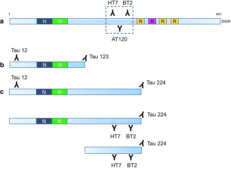
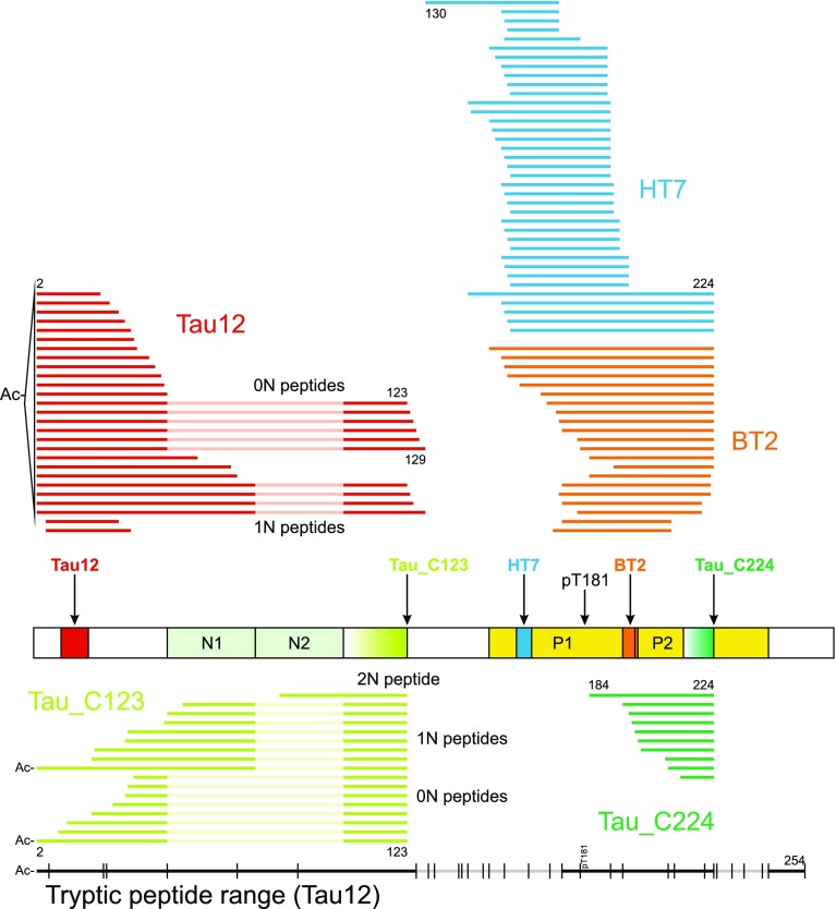
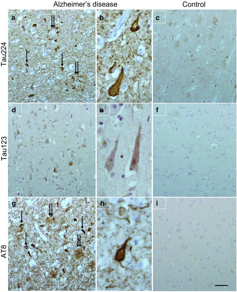
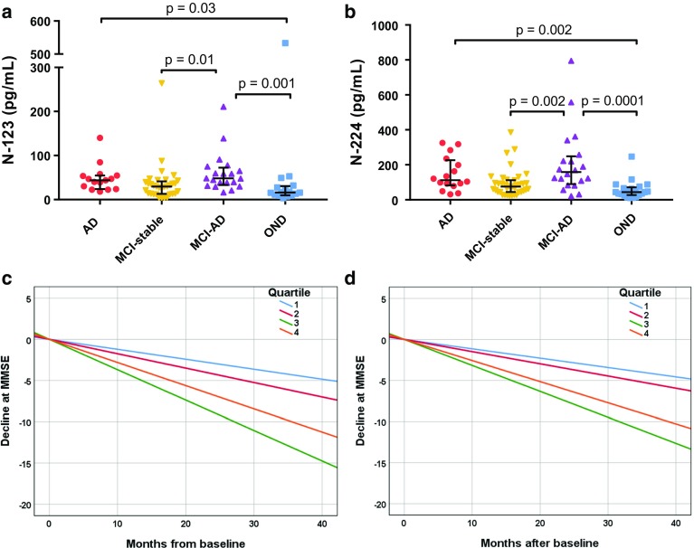
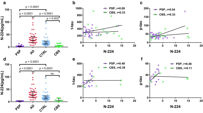
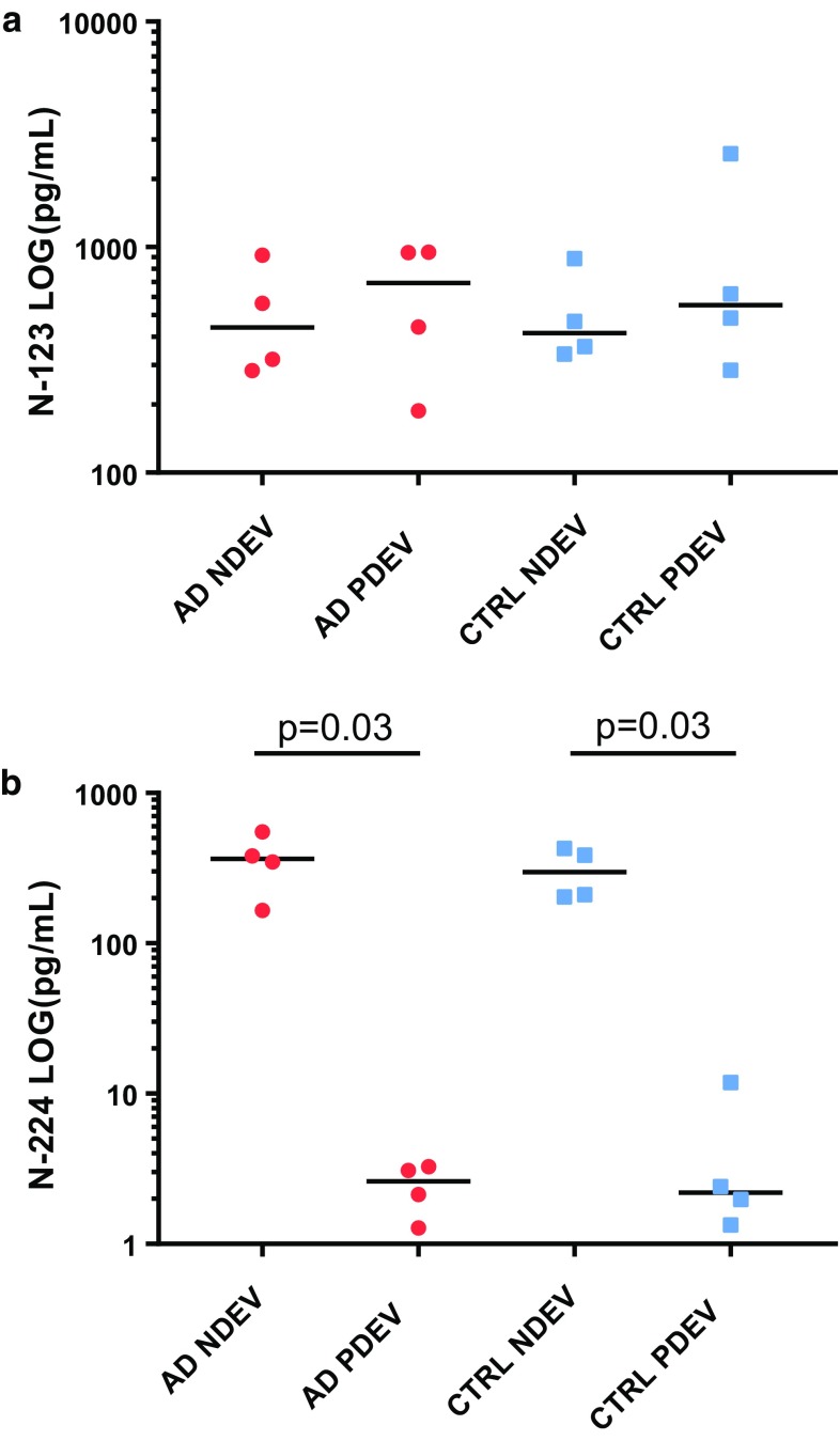
Tau is an axonal microtubule-binding protein. Tau pathology in brain and increased tau concentration in the cerebrospinal fluid (CSF) are hallmarks of Alzheimer's disease (AD). Most of tau in CSF is present as fragments. We immunoprecipitated tau from CSF and identified several endogenous peptides ending at amino acid (aa) 123 or 224 using high-resolution mass spectrometry. We raised neo-epitope-specific antibodies against tau fragments specifically ending at aa 123 and 224, respectively. With these antibodies, we performed immunohistochemistry on brain tissue and designed immunoassays measuring N-123, N-224, and x-224 tau. Immunoassays were applied to soluble brain fractions from pathologically confirmed subjects (81 AD patients, 33 controls), CSF from three cross-sectional and two longitudinal cohorts (a total of 133 AD, 38 MCI, 20 MCI-AD, 31 PSP, 15 CBS patients, and 91 controls), and neuronally- and peripherally-derived extracellular vesicles (NDEVs and PDEVs, respectively) in serum from four AD patients and four controls. Anti-tau 224 antibody stained neurofibrillary tangles and neuropil threads, while anti-tau 123 only showed weak cytoplasmic staining in AD. N-224 tau was lower in the AD soluble brain fraction compared to controls, while N-123 tau showed similar levels. N-224 tau was higher in AD compared to controls in all CSF cohorts (p < 0.001), but not N-123 tau. Decrease in cognitive performance and conversion from MCI to AD were associated with increased baseline CSF levels of N-224 tau (p < 0.0001). N-224 tau concentrations in PSP and CBS were significantly lower than in AD (p < 0.0001) and did not correlate to t-tau and p-tau. In a longitudinal cohort, CSF N-224 tau levels were stable over 6 months, with no significant effect of treatment with AChE inhibitors. N-224 tau was present in NDEVs, while N-123 tau showed comparable concentrations in both vesicle types. We suggest that N-123 tau is produced both in CNS and PNS and represents a general marker of tau metabolism, while N-224 tau is neuron-specific, present in the tangles, secreted in CSF, and upregulated in AD, suggesting a link between tau cleavage and propagation, tangle pathology, and cognitive decline.

Keywords: Alzheimer’s disease; Cerebrospinal fluid; Immunohistochemistry; Mass spectrometry; Tau fragments.


Conflict of interest statement

OH has acquired research support (for the institution) from Roche, GE Healthcare, Biogen, AVID Radiopharmaceuticals, Fujirebio, and Euroimmun. In the past 2 years, he has received consultancy/speaker fees (paid to the institution) from Lilly, Roche, and Fujirebio. KW and HHC are employed at Genentech, South San Francisco, CA, USA. HZ has served at scientific advisory board meetings for Roche Diagnostics, Wave, Samumed, and CogRx, and is a co-founder of Brain Biomarker Solutions in Gothenburg AB, a GU Venture-based platform company at the University of Gothenburg. LP has served at scientific advisory board meetings for Fujirebio, IBL and Roche.

Figures

Fig. 1
Fig. 1
Overview of standard (a) and novel (b, c) immunoassays for t-tau and tau fragments: the traditional assay only detects the mid-region of tau; our novel immunoassays detect fragments ending at aa123 and aa224
Fig. 2
Fig. 2
Top: endogenous tau species found with N-terminal and mid-region-specific antibodies (Tau 12 in red, HT7 in blue, BT2 in orange) and neo-epitope-specific antibodies (Tau_C123 in lime, Tau_C224 in green) with selected amino acid positions indicated. Alignment and numbering refer to the 2N isoform; for peptides originating from the 0N and 1N isoforms, 2N sequence portions not included are dimmed. The protein schematic structure shows the respective antibody epitopes and T181. Bottom: in black, range (Ac-2-254) of tryptic peptides detected in CSF with Tau12 (regions with no coverage are dimmed). Vertical lines indicate possible tryptic cleavage sites
Fig. 3
Fig. 3
Representation of quantitative MS data of tau fragments immunoprecipitated with Tau 12, HT7, and BT2
Fig. 4
Fig. 4
Tau immunohistochemistry in Alzheimer’s disease (a and b; d and e; g and h) and a neurologically normal control (c, f, and i). Anti-tau 224 immunohistochemistry shows neurofibrillary tangles (a, arrow) and dystrophic neurites (a, double arrow) surrounded by neuropil threads. Neurofibrillary tangles show at higher magnification (b). Faint punctuate neuronal cytoplasmic staining was observed in the normal control. Neurons in AD are weakly positive for anti-tau 123 and negative in the normal control. For comparison, sequential sections were immunohistochemically stained with AT8 to show the presence of neurofibrillary tangles in AD (g, arrow), dystrophic neurites (g, double arrow), and filamentous structure of the neurofibrillary tangles (h). AT8 staining is absent in the normal control. Bar (in i) represents 50 µm in a, c, d, f, g, and i; 20 µm in b, e, and h
Fig. 5
Fig. 5
Brain levels (normalized for total protein content) of N-123 (a), N-224 (b) fragments, and t-tau (c) in Alzheimer’s disease and controls and in AD over Braak stages III–VI (d, e, f) (lines representing the median, bars representing interquartile range)
Fig. 6
Fig. 6
Validation cohort. Left column: concentration range of N-123 (a), N-224 (b), and x-224 (c) fragments in AD and controls (lines representing median; bars representing interquartile range; in a, bars removed due to the LOG scale). Right column: correlation of N-123 (d), N-224 (e), and x-224 (f) fragment to t-tau and linear regression in AD and controls
Fig. 7
Fig. 7
Longitudinal cohort. a, b Concentration range of N-123 (a) and N-224 (b) fragments in AD, MCI, and MCI-AD and OND (lines representing the median, bars representing interquartile range). c, d Disease progression over time in AD, MCI-AD and MCI cohorts, as measured by change in MMSE scores. Higher levels of CSF N-123 (a) and N-224 (b) tau at baseline are related to faster decline. N-123: Quartile (Q) 1: n = 16, Q2: n = 15, Q3: n = 15 and Q4: n = 16; N-224: Q1: n = 17, Q2: n = 17, Q3: n = 17, and Q4: n = 17
Fig. 8
Fig. 8
PSP and CBS cohort. a, d Concentration of N-224 tau in PSP and CBS (a: Aβ+ and Aβ; d: Aβ− only) compared to AD and controls from the validation cohort (lines representing the median, bars representing interquartile range). b, c, e, f Correlation to t-tau (b) and p-tau (c) and linear regression in PSP and CBS cohorts (b, c: Aβ+ and Aβ; e, f: Aβ− only)
Fig. 9
Fig. 9
Scatter dot plots of N-123 (a) and N-224 tau (b) concentrations in serum extracellular vesicles (EVs) lysates, divided in AD and control neuronally derived EVs (NDEV) and peripherally derived EVs (PDEV) samples (lines representing median)

References

    1. Amadoro G, Corsetti V, Atlante A, Florenzano F, Capsoni S, Bussani R, et al. Interaction between NH(2)-tau fragment and Abeta in Alzheimer’s disease mitochondria contributes to the synaptic deterioration. Neurobiol Aging. 2012;33(833):833-e1. doi: 10.1016/j.neurobiolaging.2011.08.001. - DOI - PubMed
    1. Amadoro G, Corsetti V, Florenzano F, Atlante A, Ciotti MT, Mongiardi MP, et al. AD-linked, toxic NH2 human tau affects the quality control of mitochondria in neurons. Neurobiol Dis. 2014;62:489–507. doi: 10.1016/j.nbd.2013.10.018. - DOI - PubMed
    1. Amadoro G, Corsetti V, Sancesario GM, Lubrano A, Melchiorri G, Bernardini S, et al. Cerebrospinal fluid levels of a 20–22 kDa NH2 fragment of human tau provide a novel neuronal injury biomarker in Alzheimer’s disease and other dementias. J Alzheimer’s Dis. 2014;42:211–226. doi: 10.3233/JAD-140267. - DOI - PubMed
    1. Amadoro G, Corsetti V, Stringaro A, Colone M, D’Aguanno S, Meli G, et al. A NH2 tau fragment targets neuronal mitochondria at AD synapses: possible implications for neurodegeneration. J Alzheimer’s Dis. 2010;21:445–470. doi: 10.3233/JAD-2010-100120. - DOI - PubMed
    1. Amadoro G, Serafino AL, Barbato C, Ciotti MT, Sacco A, Calissano P, et al. Role of N-terminal tau domain integrity on the survival of cerebellar granule neurons. Cell Death Differ. 2004;11:217–230. doi: 10.1038/sj.cdd.4401314. - DOI - PubMed

Publication types

MeSH terms