

Site-specific 2D IR spectroscopy: a general approach for the characterization of protein dynamics with high spatial and temporal resolution
- PMID: 30548035
- PMCID: PMC6360950
- DOI: 10.1039/c8cp06146g
Site-specific 2D IR spectroscopy: a general approach for the characterization of protein dynamics with high spatial and temporal resolution
Abstract
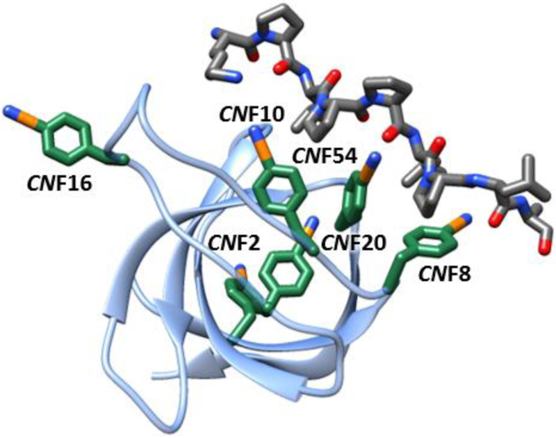
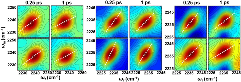
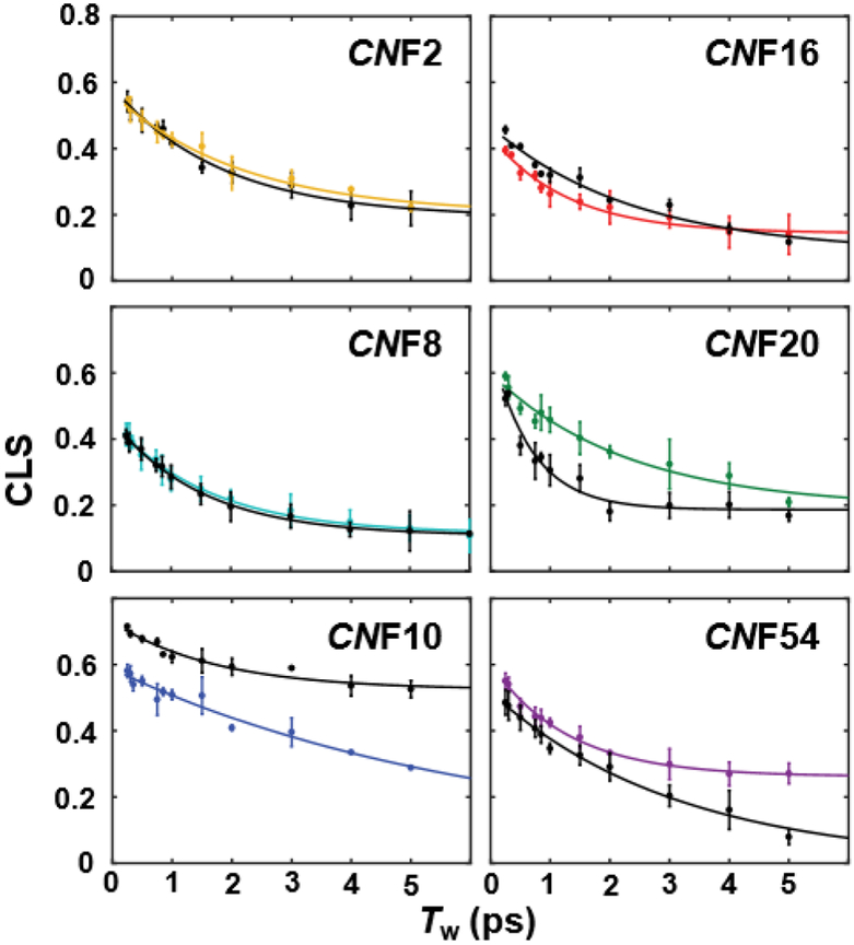
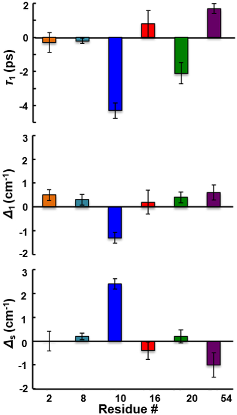
The conformational heterogeneity and dynamics of protein side chains contribute to function, but investigating exactly how is hindered by experimental challenges arising from the fast timescales involved and the spatial heterogeneity of protein structures. The potential of two-dimensional infrared (2D IR) spectroscopy for measuring conformational heterogeneity and dynamics with unprecedented spatial and temporal resolution has motivated extensive effort to develop amino acids with functional groups that have frequency-resolved absorptions to serve as probes of their protein microenvironments. We demonstrate the full advantage of the approach by selective incorporation of the probe p-cyanophenylalanine at six distinct sites in a Src homology 3 domain and the application of 2D IR spectroscopy to site-specifically characterize heterogeneity and dynamics and their contribution to cognate ligand binding. The approach revealed a wide range of microenvironments and distinct responses to ligand binding, including at the three adjacent, conserved aromatic residues that form the recognition surface of the protein. Molecular dynamics simulations performed for all the labeled proteins provide insight into the underlying heterogeneity and dynamics. Similar application of 2D IR spectroscopy and site-selective probe incorporation will allow for the characterization of heterogeneity and dynamics of other proteins, how heterogeneity and dynamics are affected by solvation and local structure, and how they might contribute to biological function.
Conflict of interest statement
Conflicts of interest
The authors declare no conflict of interest.
Figures
References
-
- Palmer AG III, Annu. Rev. Biophys. Biomol. Struct, 2001, 30, 129–155. - PubMed
MeSH terms
Substances
Grants and funding
LinkOut - more resources
Full Text Sources
Miscellaneous