

Multidelay multiparametric arterial spin labeling perfusion MRI and mild cognitive impairment in early stage Parkinson's disease
- PMID: 30548099
- PMCID: PMC6865659
- DOI: 10.1002/hbm.24451
Multidelay multiparametric arterial spin labeling perfusion MRI and mild cognitive impairment in early stage Parkinson's disease
Abstract
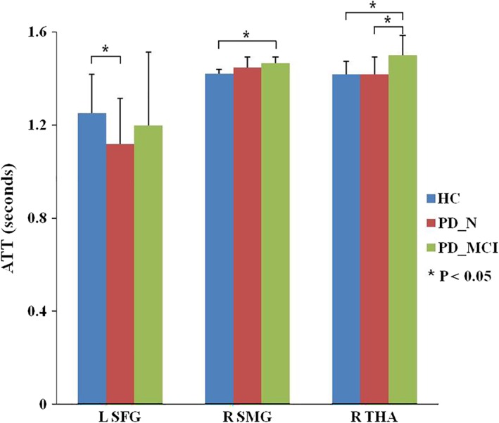
Mild cognitive impairment (MCI), a well-defined nonmotor manifestation of Parkinson's disease (PD), greatly impairs functioning and quality of life. However, the contribution of cerebral perfusion, quantified by arterial spin labeling (ASL), to MCI in PD remains poorly understood. The selection of an optimal delay time is difficult for single-delay ASL, a problem which is avoided by multidelay ASL. This study uses a multidelay multiparametric ASL to investigate cerebral perfusion including cerebral blood flow (CBF) and arterial transit time (ATT) in early stage PD patients exhibiting MCI using a voxel-based brain analysis. Magnetic resonance imaging data were acquired on a 3.0 T system at rest in 39 early stage PD patients either with MCI (PD-MCI, N = 22) or with normal cognition (PD-N, N = 17), and 36 age- and gender-matched healthy controls (HCs). CBF and ATT were compared among the three groups with SPM using analysis of variance followed by post hoc analyses to define regional differences and examine their relationship to clinical data. PD-MCI showed prolonged ATT in right thalamus compared to both PD-N and HC, and in right supramarginal gyrus compared to HC. PD-N showed shorter ATT in left superior frontal cortex compared to HC. Prolonged ATT in right thalamus was negatively correlated with the category fluency test (p = .027, r = -0.495) in the PD-MCI group. This study shows that ATT may be a more sensitive marker than CBF for the MCI, and highlights the potential role of thalamus and inferior parietal region for MCI in early stage PD.
Keywords: Parkinson's disease; arterial spin labeling; arterial transit time; cerebral blood flow; mild cognitive impairment; multidelay.
© 2018 Wiley Periodicals, Inc.
Conflict of interest statement
The authors declare no potential conflict of interests.
Figures


Similar articles
-
Changes of brain structure in Parkinson's disease patients with mild cognitive impairment analyzed via VBM technology.Neurosci Lett. 2017 Sep 29;658:121-132. doi: 10.1016/j.neulet.2017.08.028. Epub 2017 Aug 18. Neurosci Lett. 2017. PMID: 28823894
-
The cerebral blood flow deficits in Parkinson's disease with mild cognitive impairment using arterial spin labeling MRI.J Neural Transm (Vienna). 2020 Sep;127(9):1285-1294. doi: 10.1007/s00702-020-02227-6. Epub 2020 Jul 6. J Neural Transm (Vienna). 2020. PMID: 32632889
-
Discriminative pattern of reduced cerebral blood flow in Parkinson's disease and Parkinsonism-Plus syndrome: an ASL-MRI study.BMC Med Imaging. 2020 Jul 13;20(1):78. doi: 10.1186/s12880-020-00479-y. BMC Med Imaging. 2020. PMID: 32660445 Free PMC article.
-
Multidelay ASL of the pediatric brain.Br J Radiol. 2022 Jun 1;95(1134):20220034. doi: 10.1259/bjr.20220034. Epub 2022 May 12. Br J Radiol. 2022. PMID: 35451851 Free PMC article. Review.
-
Aberrant pattern of regional cerebral blood flow in mild cognitive impairment: A meta-analysis of arterial spin labeling magnetic resonance imaging.Front Aging Neurosci. 2022 Sep 1;14:961344. doi: 10.3389/fnagi.2022.961344. eCollection 2022. Front Aging Neurosci. 2022. PMID: 36118708 Free PMC article.
Cited by
-
Abnormalities of cerebral blood flow and the regional brain function in Parkinson's disease: a systematic review and multimodal neuroimaging meta-analysis.Front Neurol. 2023 Dec 7;14:1289934. doi: 10.3389/fneur.2023.1289934. eCollection 2023. Front Neurol. 2023. PMID: 38162449 Free PMC article.
-
Topologically convergent and divergent morphological gray matter networks in early-stage Parkinson's disease with and without mild cognitive impairment.Hum Brain Mapp. 2021 Oct 15;42(15):5101-5112. doi: 10.1002/hbm.25606. Epub 2021 Jul 28. Hum Brain Mapp. 2021. PMID: 34322939 Free PMC article.
-
Arterial spin labeling detects trapped labeled spins in flow-diverted aneurysms and it reflects intra-aneurysmal flow stasis.Interv Neuroradiol. 2024 Sep 27:15910199241286130. doi: 10.1177/15910199241286130. Online ahead of print. Interv Neuroradiol. 2024. PMID: 39327948 Free PMC article.
-
Reduced and Delayed Cerebrovascular Reactivity in Patients with Parkinson's Disease.Mov Disord. 2023 Jul;38(7):1262-1272. doi: 10.1002/mds.29429. Epub 2023 May 8. Mov Disord. 2023. PMID: 37157056 Free PMC article.
-
Global Alterations of Whole Brain Structural Connectome in Parkinson's Disease: A Meta-analysis.Neuropsychol Rev. 2023 Dec;33(4):783-802. doi: 10.1007/s11065-022-09559-y. Epub 2022 Sep 20. Neuropsychol Rev. 2023. PMID: 36125651 Free PMC article. Review.
References
-
- Alexander, G. E. , DeLong, M. R. , & Strick, P. L. (1986). Parallel organization of functionally segregated circuits linking basal ganglia and cortex. Annual Review of Neuroscience, 9, 357–381. - PubMed
-
- Amboni, M. , Tessitore, A. , Esposito, F. , Santangelo, G. , Picillo, M. , Vitale, C. , … Barone, P. (2015). Resting‐state functional connectivity associated with mild cognitive impairment in Parkinson's disease. Journal of Neurology, 262, 425–434. - PubMed
-
- Barzgari, A. , Sojkova, J. , Maritza Dowling, N. , Pozorski, V. , Okonkwo, O. C. , Starks, E. J. , … Gallagher, C. L. (2018). Arterial spin labeling reveals relationships between resting cerebral perfusion and motor learning in Parkinson's disease. Brain Imaging and Behaviour. 10.1007/s11682-018-9877-1 - DOI - PMC - PubMed
Publication types
MeSH terms
Substances
Grants and funding
- 81621003/National Natural Science Foundation of China/International
- 81761128023, 81220108013, 81227002, 81030027/National Natural Science Foundation of China/International
- IRT16R52/Program for Changjiang Scholars and Innovative Research Team in University of China/International
- T2014190/Changjiang Scholar Professorship Award of China/International
- F510000/G16916411/CMB Distinguished Professorship Award administered by the Institute of International Education/International
LinkOut - more resources
Full Text Sources
Medical
