Review
. 2019 Jul;26(5):e12520.
doi: 10.1111/micc.12520. Epub 2019 Jan 17.

Methods to label, image, and analyze the complex structural architectures of microvascular networks

Affiliations
Review

Methods to label, image, and analyze the complex structural architectures of microvascular networks

Bruce A Corliss et al. Microcirculation. 2019 Jul.

Abstract

Microvascular networks play key roles in oxygen transport and nutrient delivery to meet the varied and dynamic metabolic needs of different tissues throughout the body, and their spatial architectures of interconnected blood vessel segments are highly complex. Moreover, functional adaptations of the microcirculation enabled by structural adaptations in microvascular network architecture are required for development, wound healing, and often invoked in disease conditions, including the top eight causes of death in the Unites States. Effective characterization of microvascular network architectures is not only limited by the available techniques to visualize microvessels but also reliant on the available quantitative metrics that accurately delineate between spatial patterns in altered networks. In this review, we survey models used for studying the microvasculature, methods to label and image microvessels, and the metrics and software packages used to quantify microvascular networks. These programs have provided researchers with invaluable tools, yet we estimate that they have collectively attained low adoption rates, possibly due to limitations with basic validation, segmentation performance, and nonstandard sets of quantification metrics. To address these existing constraints, we discuss opportunities to improve effectiveness, rigor, and reproducibility of microvascular network quantification to better serve the current and future needs of microvascular research.

Keywords: blood vessels; image analysis; image quantification; microvasculature; vascular network; vessel architecture.


Figures

Figure 1
Figure 1
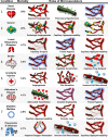
The significance of the microvasculature in top causes of death and disease in United States. Top eight classes of fatal disease or injury with the fraction of annual deaths in the United States. Included with each malady are three highlighted fundamental roles the microvasculature plays with initiation, maintenance, or treatment (see main text for references)
Figure 2
Figure 2
Both endothelial cells and pericytes share markers used in the literature for labeling endothelial cells, and Col‐IV tracks, assumed to be regressed vessels, lack a pixel intensity profile indicative of a lumen. (A) Retina capillary with pericyte (NG2, red), IB4 lectin (green), endothelial cells (CD31, yellow), and cell nuclei (DAPI, cyan). (B) Retina capillary with pericyte and endothelial cells labeled with Col‐IV (green; scale bar 10 μm). (C) High‐resolution image of Col‐IV off‐vessel track (star) and lumenized blood vessel (arrow; scale bar 5 μm). (D) Comparison of Col‐IV relative pixel intensity profile across cross‐section of blood vessels and collagen tracks (P = 8.11E‐6, 2‐way analysis of variance, N = 10 vessels and tracks, error bars are standard deviation)
Figure 3
Figure 3
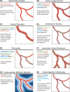
Basic metrics quantifying the complexities of the microvascular architecture. Visual explanation of metrics that have been used to quantify various aspects of microvessel network architecture, including (A) VAF, (B) vessel length density, (C) vessel diameter, (D) branchpoints density, (E) tortuosity, (F) lacranuity and fractal dimension, (G) extra‐vascular diffusion distance, and (H) vessel segment partitioning
Figure 4
Figure 4
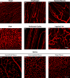
Heterogeneity of blood vessel network structure across and within tissue. IB4 lectin Perfused microvessels of (A) heart, (B) diaphragm, (C) skeletal muscle, (D) liver, (E) peritoneal cavity, (F) inguinal fat, and of the three distinct vascular layers of the retina (G‐I; scale bar 50 μm)
Figure 5
Figure 5
Bridging form and function: correspondence between microvasculature architecture metrics and biological behaviors. Schematized microvasculature network with various cellular and acellular components (multicolored font) mapped to quantitative image analysis metrics that indicate different aspects of microvascular function (black font)
Figure 6
Figure 6
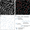
Novel image analysis metrics by analyzing the microvasculature with graph theory. A, Confocal microscopy image of the murine retinal deep vasculature, with CD105 marking ECs (white; scale bar = 50 μm). B, Image analyzed with basic segmentation, skeletonization, and branchpoints classification, with vessel centerline/skeleton (white), branchpoints (red), and EP (turquoise). C, Conversion of vasculature into a graph, with branchpoints indicated as “nodes” (blue) and vessel segments as “edges” (gray). D, Visual summary of types of metrics to quantify graphs, with removed edges representing change to network after a blood vessel has regressed or experiences obstructed flow

References

    1. Jacob M, Chappell D, Becker BF. Regulation of blood flow and volume exchange across the microcirculation. Crit Care. 2016;20:319. - PMC - PubMed
    1. Nourshargh S, Alon R. Leukocyte migration into inflamed tissues. Immunity. 2014;41(5):694‐707. - PubMed
    1. Tonnesen MG, Feng X, Clark RAF. Angiogenesis in wound healing. J Investig Dermatol Symp Proc. 2000;5(1):40‐46. - PubMed
    1. Chambers R, Zweifach BW. Intercellular cement and capillary permeability. Physiol Rev. 1947;27(3):436‐463. - PubMed
    1. Pantoni L. Cerebral small vessel disease: from pathogenesis and clinical characteristics to therapeutic challenges. Lancet Neurol. 2010;9(7):689‐701. - PubMed

Publication types