Case Reports
. 2019 Jan;179(1):94-103.
doi: 10.1002/ajmg.a.60687. Epub 2018 Dec 14.

An N-terminal heterozygous missense CASK mutation is associated with microcephaly and bilateral retinal dystrophy plus optic nerve atrophy

Affiliations
Case Reports

An N-terminal heterozygous missense CASK mutation is associated with microcephaly and bilateral retinal dystrophy plus optic nerve atrophy

Leslie E W LaConte et al. Am J Med Genet A. 2019 Jan.

Abstract

Heterozygous loss-of-function mutations in the X-linked gene CASK are associated with mental retardation and microcephaly with pontine and cerebellar hypoplasia (MICPCH) and ophthalmological disorders including optic nerve atrophy (ONA) and optic nerve hypoplasia (ONH). Recently, we have demonstrated that CASK(+/-) mice display ONH with 100% penetrance but exhibit no change in retinal lamination or structure. It is not clear if CASK loss-of-function predominantly affects retinal ganglion cells, or if other retinal cells like photoreceptors are also involved. Here, we report a heterozygous missense mutation in the N-terminal calcium/calmodulin-dependent kinase (CaMK) domain of the CASK protein in which a highly conserved leucine is mutated to the cyclic amino acid proline. In silico analysis suggests that the mutation may produce destabilizing structural changes. Experimentally, we observe pronounced misfolding and insolubility of the CASKL209P protein. Interestingly, the remaining soluble mutant protein fails to interact with Mint1, which specifically binds to CASK's CaMK domain, suggesting a mechanism for the phenotypes observed with the CASKL209P mutation. In addition to microcephaly, cerebellar hypoplasia and delayed development, the subject with the L209P mutation also presented with bilateral retinal dystrophy and ONA. Electroretinography indicated that rod photoreceptors are the most prominently affected cells. Our data suggest that the CASK interactions mediated by the CaMK domain may play a crucial role in retinal function, and thus, in addition to ONH, individuals with mutations in the CASK gene may exhibit other retinal disorders, depending on the nature of mutation.

Keywords: CASK; MICPCH; optic nerve atrophy; optic nerve hypoplasia; retinal dystrophy.


Conflict of interest statement

Conflict of interest

The authors have no conflicts of interest to report.

Figures

Figure 1.
Figure 1.. Clinical findings for subject with CASKL209P mutation.
A) Subject’s clinical presentation. B) T2-weighted MRI brain scans from subject displaying small cerebellum (white arrow) and C) optic nerve (white arrow) at age 5 years. D) Wide field images of the retina of the right (left panel) and left (right panel) eyes show optic atrophy and arteriolar attenuation but no significant pigment migration at age 7 years. E) Results from full-field electroretinography examination. The scotopic maximal response, representing both cones and rods, is electronegative and is moderately reduced in right (top) and left (bottom) eyes at 5 years. The dashed lines indicate the normal amplitude of trough and crest.
Figure 2.
Figure 2.. Impact of L209P mutation on CASK structure.
A) Schematic of the domains of CASK, including locations of known interactions with other proteins, and the site of the L209P mutation. B) Conservation of the mutated site in various animal species. C) Box and whisker plots of the radius of gyration and root mean square deviation (RMSD) of the backbone of the CAMK domain of CASK or CASKL209P over the course of three molecular dynamics (MD) trajectories, respectively. D) Structure of CASK’s CAMK domain, modeled from a crystal structure (3C0H.pdb), with a proline substituted for leucine at position 209 (red) in the structurally important αF helix (blue). Other structurally critical regions of the core protein kinase catalytic domain are also shown: αH helix (pink), R-spine (red space-filling residues), and C-spine (yellow space-filling residues). The activation segment is shown in green, with regions of increased mobility due to the L209P mutation as predicted by MD simulations shown in orange. E) Average B-factors (±SEM) for the two regions (residues 166–168 and 174–176; shown in orange in panel D) within the activation segment (green, panel D) of CASK’s CAMK domain that demonstrated notable differences between CASK and CASKL209P.
Figure 3.
Figure 3.. Properties of GFP-CASKL209P expressed in cell culture.
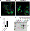
A,B) Representative images of wildtype CASK and CASKL209P GFP fusion protein expressed in HEK293 cells. Note the cotton-wool aggregates of CASKL209P evident in some cells. Scale bar= 5µm. C) Quantitation of number of cells displaying large aggregations. Results represent mean and SEM from 6 different experiments. A total of ~1000 cells were examined in each sample. D) Blot for CASK in insoluble and soluble fractions of cell lysate. CASK antibody detects both expressed GFP-CASK and endogenous CASK present in HEK cells. Note that CASKL209P is predominantly present in the insoluble fraction.
Figure 4.
Figure 4.. Neurexin-mediated recruitment assay in HEK293 cells.
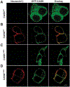
Confocal images of HEK 293 cells transfected with A) GFP-CASK alone, B) GFP-CASK with FLAG-tagged neurexin1-β, C) GFP-CASKL209P alone, and D) GFP-CASKL209P with FLAG-tagged neurexin1-β. Scale bar= 5µm
Figure 5.
Figure 5.. Pull-down assays to evaluate interactions of CASKL209P with other proteins.
A) Schematic of GST pull-down from transfected HEK293 cells. B) Schematic of immunoprecipitation assay using GFP antibody. C) Immunoblot of CASK from HEK 293 cells. Endogenous CASK can be seen in GFP sample. D) Blots of indicated antigen from immunoprecipitation assay.

References

    1. Atasoy D, Schoch S, Ho A, Nadasy KA, Liu X, Zhang W, … Sudhof TC (2007). Deletion of CASK in mice is lethal and impairs synaptic function. Proc Natl Acad Sci U S A, 104(7), 2525–2530. doi:0611003104 [pii]10.1073/pnas.0611003104 - DOI - PMC - PubMed
    1. Burglen L, Chantot-Bastaraud S, Garel C, Milh M, Touraine R, Zanni G, … Rodriguez D (2012). Spectrum of pontocerebellar hypoplasia in 13 girls and boys with CASK mutations: confirmation of a recognizable phenotype and first description of a male mosaic patient. Orphanet J Rare Dis, 7, 18. doi:1750–1172-7–18 [pii]10.1186/1750-1172-7-18 - DOI - PMC - PubMed
    1. Butz S, Okamoto M, & Sudhof TC (1998). A tripartite protein complex with the potential to couple synaptic vesicle exocytosis to cell adhesion in brain. Cell, 94(6), 773–782. - PubMed
    1. Chaplin C, Borchert MS, Fink C, Garcia-Filion P, & McCulloch DL (2009). Light-adapted electroretinograms in optic nerve hypoplasia. Doc Ophthalmol, 119(2), 123–132. doi:10.1007/s10633-009-9188-3 - DOI - PubMed
    1. Cibis GW, & Fitzgerald KM (1994). Optic nerve hypoplasia in association with brain anomalies and an abnormal electroretinogram. Doc Ophthalmol, 86(1), 11–22. - PubMed

Publication types

MeSH terms