

Relationship between Progressive Changes in Lamina Cribrosa Depth and Deterioration of Visual Field Loss in Glaucomatous Eyes
- PMID: 30549470
- PMCID: PMC6288017
- DOI: 10.3341/kjo.2018.0015
Relationship between Progressive Changes in Lamina Cribrosa Depth and Deterioration of Visual Field Loss in Glaucomatous Eyes
Abstract
Purpose: To investigate the relationship between the progression of visual field (VF) loss and changes in lamina cribrosa depth (LCD) as determined by spectral-domain optical coherence tomography (SD-OCT) enhanced depth imaging in patients with primary open angle glaucoma (POAG).
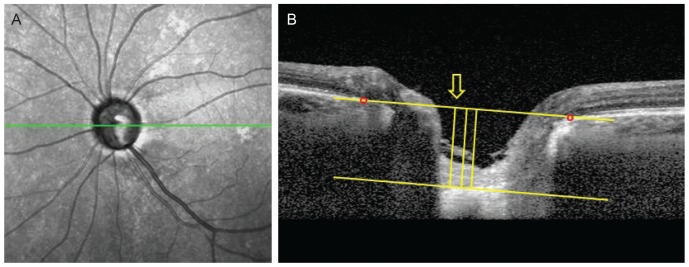
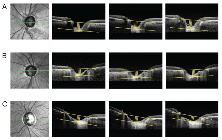
Methods: Data from 60 POAG patients (mean follow-up, 3.5 ± 0.7 years) were included in this retrospective study. The LCD was measured in the optic disc image using SD-OCT enhanced depth imaging scanning at each visit. Change in the LCD was considered to either 'increase' or 'decrease' when the differences between baseline and the latest two consecutive follow-up visits were greater than the corresponding reproducibility coefficient value (23.08 μm, as determined in a preliminary reproducibility study). All participants were divided into three groups: increased LCD (ILCD), decreased LCD (DLCD), and no LCD change (NLCD). The Early Manifest Glaucoma Trial criteria were used to define VF deterioration. Kaplan-Meier survival analysis and Cox's proportional hazard models were performed to explore the relationship between VF progression and LCD change.
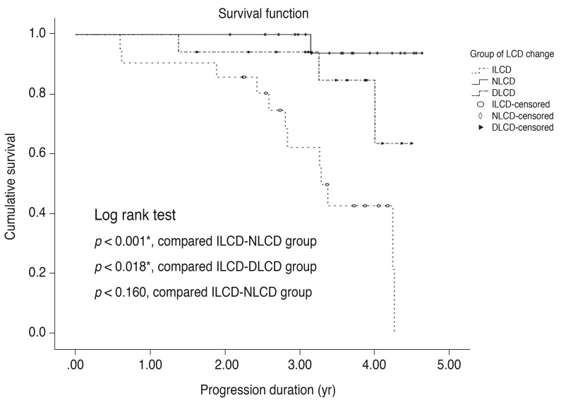
Results: Of the 60 eyes examined, 35.0% (21 eyes), 28.3% (17 eyes), and 36.7% (22 eyes) were classified as the ILCD, DLCD, and NLCD groups, respectively. Kaplan-Meier survival analysis showed a greater cumulative probability of VF progression in the ILCD group than in the NLCD (p < 0.001) or DLCD groups (p = 0.018). Increased LCD was identified as the only risk factor for VF progression in the Cox proportional hazard models (hazard ratio, 1.008; 95% confidence interval, 1.000 to 1.015; p = 0.047).
Conclusions: Increased LCD was associated with a greater possibility of VF progression. The quantitative measurement of LCD changes, determined by SD-OCT, is a potential biomarker for the prediction of VF deterioration in patients with POAG.
Keywords: Glaucoma; Lamina cribrosa; Optic disk; Optical coherence tomography; Visual fields.
© 2018 The Korean Ophthalmological Society.
Conflict of interest statement
No potential conflict of interest relevant to this article was reported.
Figures
Similar articles
-
Baseline Lamina Cribrosa Curvature and Subsequent Visual Field Progression Rate in Primary Open-Angle Glaucoma.Ophthalmology. 2018 Dec;125(12):1898-1906. doi: 10.1016/j.ophtha.2018.05.017. Epub 2018 Jun 23. Ophthalmology. 2018. PMID: 29945800
-
Effect of focal lamina cribrosa defect on glaucomatous visual field progression.Ophthalmology. 2014 Aug;121(8):1524-30. doi: 10.1016/j.ophtha.2014.02.017. Epub 2014 Mar 31. Ophthalmology. 2014. PMID: 24697910
-
Association of Functional Loss With the Biomechanical Response of the Optic Nerve Head to Acute Transient Intraocular Pressure Elevations.JAMA Ophthalmol. 2018 Feb 1;136(2):184-192. doi: 10.1001/jamaophthalmol.2017.6111. JAMA Ophthalmol. 2018. PMID: 29302683 Free PMC article.
-
Microstructure of Peripapillary Atrophy and Subsequent Visual Field Progression in Treated Primary Open-Angle Glaucoma.Ophthalmology. 2016 Mar;123(3):542-51. doi: 10.1016/j.ophtha.2015.10.061. Epub 2015 Dec 12. Ophthalmology. 2016. PMID: 26692299
-
Glymphatic stasis at the site of the lamina cribrosa as a potential mechanism underlying open-angle glaucoma.Clin Exp Ophthalmol. 2017 Jul;45(5):539-547. doi: 10.1111/ceo.12915. Epub 2017 Feb 27. Clin Exp Ophthalmol. 2017. PMID: 28129671 Review.
Cited by
-
Discovery and clinical translation of novel glaucoma biomarkers.Prog Retin Eye Res. 2021 Jan;80:100875. doi: 10.1016/j.preteyeres.2020.100875. Epub 2020 Jul 10. Prog Retin Eye Res. 2021. PMID: 32659431 Free PMC article. Review.
-
Racial Differences in the Rate of Change in Anterior Lamina Cribrosa Surface Depth in the African Descent and Glaucoma Evaluation Study.Invest Ophthalmol Vis Sci. 2021 Apr 1;62(4):12. doi: 10.1167/iovs.62.4.12. Invest Ophthalmol Vis Sci. 2021. PMID: 33844828 Free PMC article.
References
-
- Quigley HA, Addicks EM, Green WR, Maumenee AE. Optic nerve damage in human glaucoma. II. The site of injury and susceptibility to damage. Arch Ophthalmol. 1981;99:635–649. - PubMed
-
- Quigley HA, Anderson DR. Distribution of axonal transport blockade by acute intraocular pressure elevation in the primate optic nerve head. Invest Ophthalmol Vis Sci. 1977;16:640–644. - PubMed
-
- Radius RL, Anderson DR. Rapid axonal transport in primate optic nerve. Distribution of pressure-induced interruption. Arch Ophthalmol. 1981;99:650–654. - PubMed
MeSH terms
LinkOut - more resources
Full Text Sources
Medical
