. 2019 Jan;38 Suppl 1(Iss 1 Suppl 1):S9-S24.
doi: 10.1097/PGP.0000000000000552.

Endometrial Carcinoma, Grossing and Processing Issues: Recommendations of the International Society of Gynecologic Pathologists

Affiliations

Endometrial Carcinoma, Grossing and Processing Issues: Recommendations of the International Society of Gynecologic Pathologists

Anais Malpica et al. Int J Gynecol Pathol. 2019 Jan.

Abstract

Endometrial cancer is the most common gynecologic neoplasm in developed countries; however, updated universal guidelines are currently not available to handle specimens obtained during the surgical treatment of patients affected by this disease. This article presents recommendations on how to gross and submit sections for microscopic examination of hysterectomy specimens and other tissues removed during the surgical management of endometrial cancer such as salpingo-oophorectomy, omentectomy, and lymph node dissection-including sentinel lymph nodes. In addition, the intraoperative assessment of some of these specimens is addressed. These recommendations are based on a review of the literature, grossing manuals from various institutions, and a collaborative effort by a subgroup of the Endometrial Cancer Task Force of the International Society of Gynecological Pathologists. The aim of these recommendations is to standardize the processing of endometrial cancer specimens which is vital for adequate pathological reporting and will ultimately improve our understanding of this disease.


Conflict of interest statement

The authors declare no conflict of interest.

Figures

FIG. 1
FIG. 1
Orientation of hysterectomy specimen using anatomical landmarks, peritoneal reflection is higher anteriorly (arrow) and the sequence of structures in the adnexal region is round ligament (*), fallopian tube (arrowhead) and ovary (**) (A), peritoneal reflection is lower posteriorly (arrow) and the sequence of structures in the adnexal region is ovary (**), fallopian tube (arrowhead) and round ligament (B), the latter is not visualized in this photograph.
FIG. 2
FIG. 2
Inking the anterior and posterior uterine serosal surfaces with extension of the ink to the ectocervix or vaginal cuff margin (A), helps to confirm the presence of tumor in the uterine serosa (arrow) (B), and in cases with cervical stromal involvement (C) the measurement of the relationship of the stromal invasion to the full thickness of the cervical wall (line), and the proper identification of the ectocervical or vaginal cuff margin (*) (D).
FIG. 3
FIG. 3
Opening the uterus at 3 and 9 o’clock provides a maximum exposure of the endometrial surface in a flat plane to better visualize and measure an endometrial tumor (A), the cornua can contain tumor (B) and this should not be mistaken for myometrial invasion (C).
FIG. 4
FIG. 4
Measurement of endometrial carcinoma, small polypoid tumor (A), a tumor that involves the anterior and posterior walls in a continuum, carpet-like fashion should be measured accordingly (B).
FIG. 5
FIG. 5
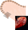
Cross-sections of the uterine wall at the level of the corpus (top arrow), longitudinal sections at the level of the lower uterine segment (middle arrow) and cervix (bottom arrow).
FIG. 6
FIG. 6
Cross-section of the uterine wall demonstrating deepest point of myometrial invasion on gross examination (arrowhead).
FIG. 7
FIG. 7
Sectioning and Extensively Examining the FIMbriated End protocol, sectioning of adnexa to be submitted for microscopic examination.
FIG. 8
FIG. 8
Sentinel lymph node, parallel slices are perpendicular to the long axis of the specimen.
FIG. 9
FIG. 9
Ultrastaging protocols, MD Anderson Cancer Center (MDACC) (A) and Memorial Sloan Kettering Cancer Center (MSKCC), ultrastaging will be obtained if there is myometrial or vascular/lymphatic invasion (*) (B). H&E indicates hematoxylin and eosin; IHC, immunohistochemistry; SLN, sentinel lymph node.

References

    1. Lortet-Tieulent J, Ferlay J, Bray F, et al. International patterns and trends in endometrial cancer incidence, 1978-2013. J Natl Cancer Inst 2017;110:354–361. - PubMed
    1. American Cancer Society. Cancer Facts & Figures. Altanta CA: American Cancer Society; 2018.
    1. Houghton JP, Roddy S, Carroll S, et al. A simple method for the prevention of endometrial autolysis in hysterectomy specimens. J Clin Pathol 2004;57:332–3. - PMC - PubMed
    1. Hicks DG, Boyce BF. The challenge and importance of standardizing pre-analytical variables in surgical pathology specimens for clinical care and translational research. Biotech Histochem 2012;87:14–7. - PubMed
    1. Khoury T. Delay to formalin fixation alters morphology and immunohistochemistry for breast carcinoma. Appl Immunohistochem Mol Morphol 2012;20:531–42. - PubMed