. 2018 Dec 14;12(12):e0007011.
doi: 10.1371/journal.pntd.0007011. eCollection 2018 Dec.

A high throughput neutralization test based on GFP expression by recombinant rabies virus

Affiliations

A high throughput neutralization test based on GFP expression by recombinant rabies virus

Jillybeth Burgado et al. PLoS Negl Trop Dis. .

Abstract

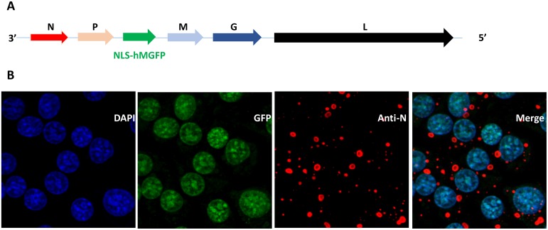
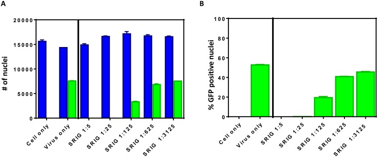
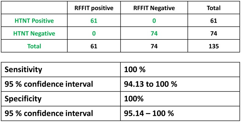
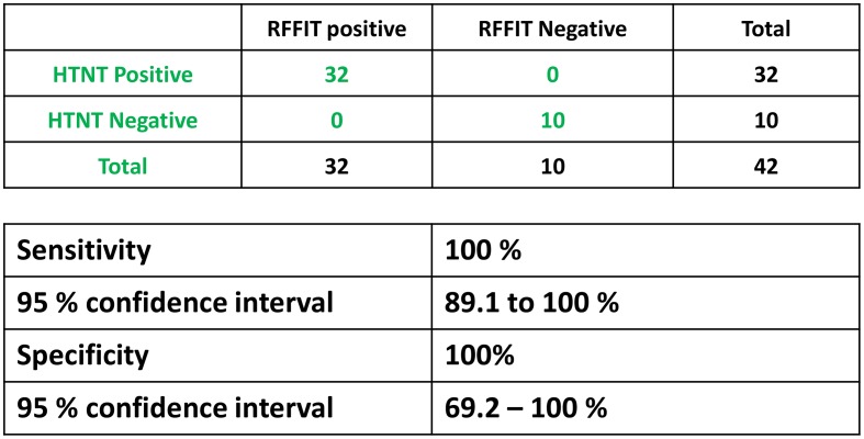
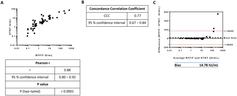
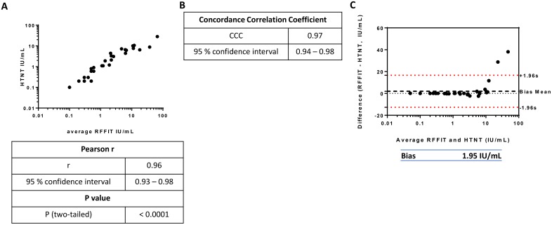
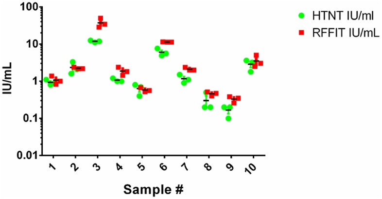
The effectiveness of rabies vaccination in both humans and animals is determined by the presence of virus neutralizing antibodies (VNAs). The Rapid Fluorescent Focus Inhibition Test (RFFIT) is the method traditionally used for detection and quantification of VNAs. It is a functional in vitro test for assessing the ability of antibodies in serum to bind and prevent infection of cultured cells with rabies virus (RABV). The RFFIT is a labor intensive, low throughput and semi-quantitative assay performed by trained laboratorians. It requires staining of RABV-infected cells by rabies specific fluorescent antibodies and manual quantification of fluorescent fields for titer determination. Although the quantification of fluorescent fields observed in each sample is recorded, the corresponding images are not stored or captured to be used for future analysis. To circumvent several of these disadvantages, we have developed an alternative, automated high throughput neutralization test (HTNT) for determination of rabies VNAs based on green fluorescent protein (GFP) expression by a recombinant RABV and compared with the RFFIT. The HTNT assay utilizes the recombinant RABV ERA variant expressing GFP with a nuclear localization signal (NLS) for efficient quantification. The HTNT is a quantitative method where the number of RABV-infected cells are determined and the images are stored for future analysis. Both RFFIT and HTNT results correlated 100% for a panel of human and animal positive and negative rabies serum samples. Although, the VNA titer values are generally agreeable, HTNT titers tend to be lower than that of RFFIT, probably due to the differences in quantification methods. Our data demonstrates the potential for HTNT assays in determination of rabies VNA titers.


Conflict of interest statement

The authors have declared that no competing interests exist.

Figures

Fig 1
Fig 1. Recombinant RABV ERA-NLS-hMGFP.
(A) The schematic representation of recombinant RABV ERA genome with the inserted NLS-hMGFP. 3’ and 5’ denotes the direction of negative sense genome. N, P, M, G and L corresponds to nucleoprotein, phosphoprotein, matrix protein, glycoprotein and large RNA dependent RNA polymerase genes, respectively. (B) BSR cells were infected with recombinant RABV ERA-NLS-hMGFP virus for 24 h at 37°C. The cells were stained with DAPI (blue) and anti-N monoclonal antibodies (red) and imaged. The merge represents the co-localization of NLS-hMGFP with DAPI demonstrating nuclear targeting of GFP (green).
Fig 2
Fig 2. Quantification of DAPI and GFP positive cells by HTNT.
BSR cells were infected with ERA-NLS-GFP for 20 h at 37°C in the presence and absence of SRIG and stained with DAPI (for nucleus). The number of DAPI and GFP positive nuclei are quantitated. (A) Total number of DAPI stained nuclei (corresponding to number of cells) and GFP co-localization with DAPI stained cells are shown as blue and green bars, respectively. (B) The percent GFP positive cells under various conditions were calculated.
Fig 3
Fig 3. Sensitivity and specificity of human sera.
Neutralization capability of rabies-specific antibodies in sera from vaccinated and naïve individuals was assessed using RFFIT and HTNT. Based on the complete neutralization of RABV at the 1:5 serum dilution samples were classified as positive or negative. Sensitivity and specificity of HTNT assay for detection of rabies VNAs were calculated based on the RFFIT results and tabulated with 95% confidence interval levels.
Fig 4
Fig 4. Sensitivity and specificity of animal sera.
Neutralization capability of rabies-specific antibodies in animal determined similar to Fig 4. Sensitivity and specificity of HTNT assay for detection of rabies VNAs were calculated based on the RFFIT results and tabulated with 95% confidence interval levels.
Fig 5
Fig 5. Comparison of HTNT and RFFIT titers of human sera.
Titers (IU/ml) of positive human sera were compared between the two methods. Titers were log-transformed for normalization purposes. (A) HTNT titers were plotted against RFFIT titers to determine the Pearson correlation coefficient. Similarly, the concordance correlation coefficient (B) and the Bland-Altman plot of differences (C) were calculated. In (C), the mean difference of the titers is represented by the Bias value, plotted as a black dotted line above 0. The limits of agreement, within which 95% of the differences between RFFIT and HTNT are denoted by the red dotted lines above and below 1.96 standard deviations of the mean difference.
Fig 6
Fig 6. Comparison of HTNT and RFFIT titers of animal sera.
The statistical analysis of animal sera were performed similar to Fig 6. The Pierson correlation coefficient (A), concordance correlation coefficient (B) and the Bland-Altman plot to determine bias value of titers (IU/ml) of positive animal sera (C) are provided. In (C) the mean difference of the titers is represented by the Bias value, plotted as a black dotted line above 0. The limits of agreement, within which 95% of the differences between RFFIT and HTNT are denoted by the red dotted lines above and below 1.96 standard deviations of the mean difference.
Fig 7
Fig 7. Reproducibility of HTNT assay.
A subset of animal serum samples (n = 14) were run in triplicate on different days using both methods. Negative sera did not neutralize at the 1:5 dilution in either assay. Individual IU/ml titers for positive samples are plotted to evaluate the inter-assay variability.

References

    1. Fooks AR, Banyard AC, Horton DL, Johnson N, McElhinney LM, Jackson AC. Current status of rabies and prospects for elimination. Lancet. 2014;384(9951):1389–99. Epub 2014/05/16. 10.1016/S0140-6736(13)62707-5 . - DOI - PMC - PubMed
    1. Moore SM, Hanlon CA. Rabies-specific antibodies: measuring surrogates of protection against a fatal disease. PLoS Negl Trop Dis. 2010;4(3):e595 Epub 2010/03/17. 10.1371/journal.pntd.0000595 . - DOI - PMC - PubMed
    1. Cox JH, Dietzschold B, Schneider LG. Rabies virus glycoprotein. II. Biological and serological characterization. Infect Immun. 1977;16(3):754–9. Epub 1977/06/01. . - PMC - PubMed
    1. Warrell MJ. Current rabies vaccines and prophylaxis schedules: preventing rabies before and after exposure. Travel Med Infect Dis. 2012;10(1):1–15. Epub 2012/02/22. 10.1016/j.tmaid.2011.12.005 . - DOI - PubMed
    1. Manning SE, Rupprecht CE, Fishbein D, Hanlon CA, Lumlertdacha B, Guerra M, et al. Human rabies prevention—United States, 2008: recommendations of the Advisory Committee on Immunization Practices. MMWR Recomm Rep. 2008;57(RR-3):1–28. Epub 2008/05/23. . - PubMed

Publication types

MeSH terms