. 2018 Dec 13;175(7):1756-1768.e17.
doi: 10.1016/j.cell.2018.10.025.

Irisin Mediates Effects on Bone and Fat via αV Integrin Receptors

Affiliations

Irisin Mediates Effects on Bone and Fat via αV Integrin Receptors

Hyeonwoo Kim et al. Cell. .

Erratum in

  • Irisin Mediates Effects on Bone and Fat via αV Integrin Receptors.
    Kim H, Wrann CD, Jedrychowski M, Vidoni S, Kitase Y, Nagano K, Zhou C, Chou J, Parkman VA, Novick SJ, Strutzenberg TS, Pascal BD, Le PT, Brooks DJ, Roche AM, Gerber KK, Mattheis L, Chen W, Tu H, Bouxsein ML, Griffin PR, Baron R, Rosen CJ, Bonewald LF, Spiegelman BM. Kim H, et al. Cell. 2019 Jul 11;178(2):507-508. doi: 10.1016/j.cell.2019.06.028. Cell. 2019. PMID: 31299203 Free PMC article. No abstract available.

Abstract

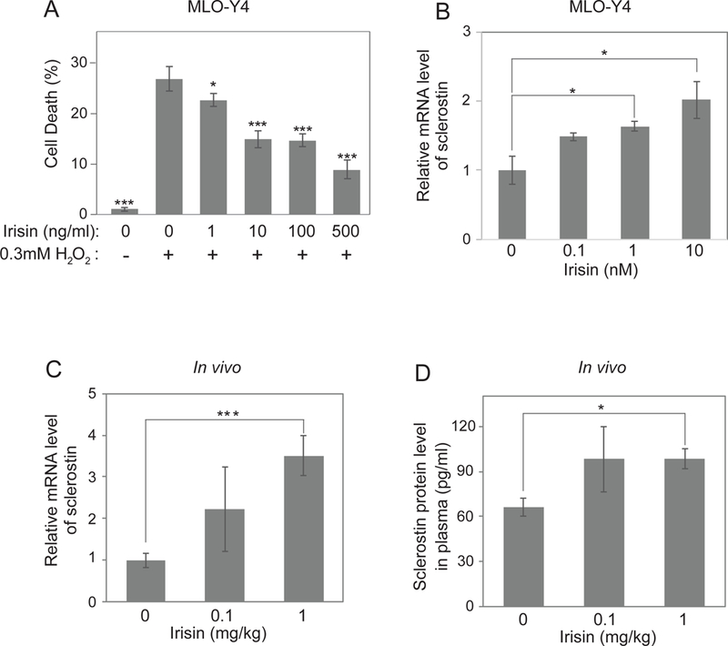
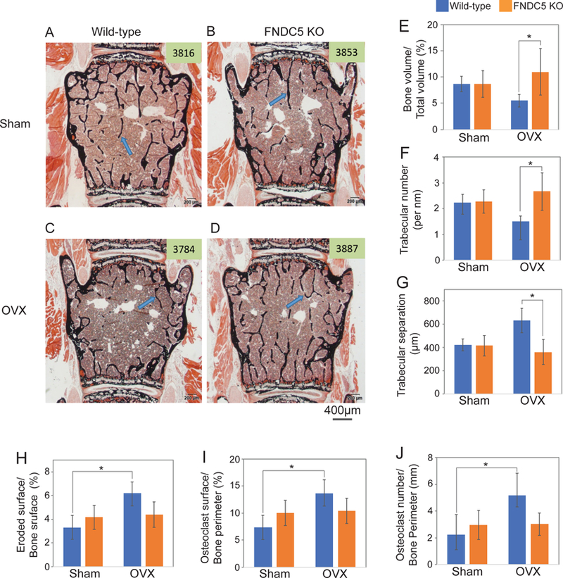
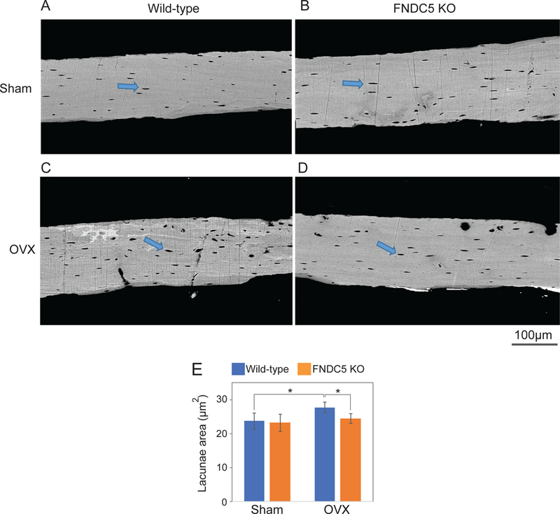
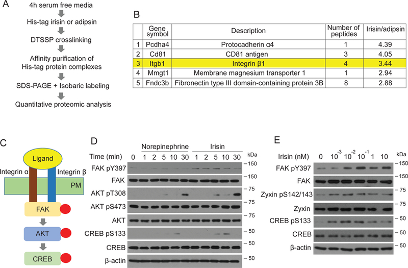
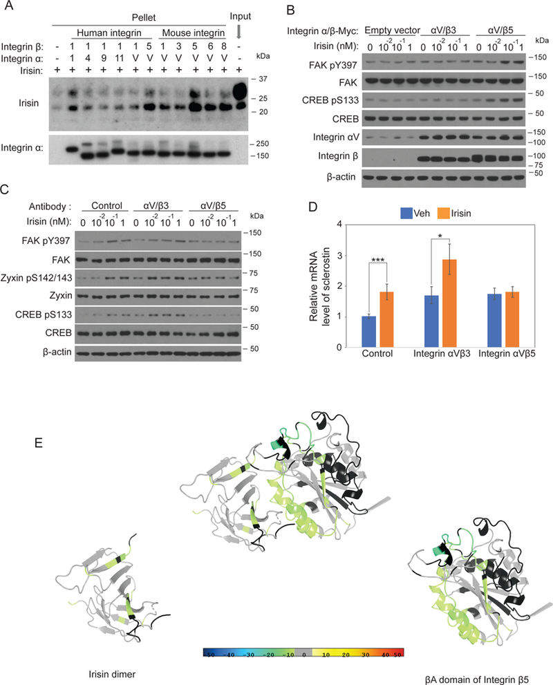
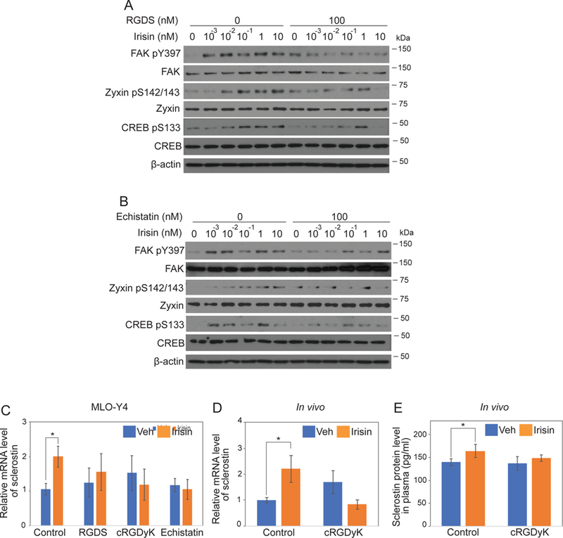
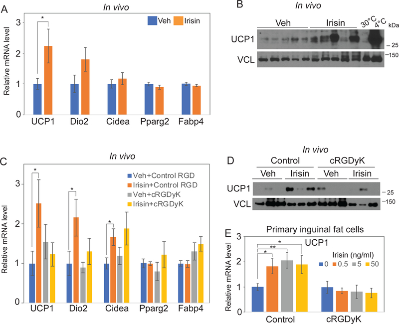
Irisin is secreted by muscle, increases with exercise, and mediates certain favorable effects of physical activity. In particular, irisin has been shown to have beneficial effects in adipose tissues, brain, and bone. However, the skeletal response to exercise is less clear, and the receptor for irisin has not been identified. Here we show that irisin binds to proteins of the αV class of integrins, and biophysical studies identify interacting surfaces between irisin and αV/β5 integrin. Chemical inhibition of the αV integrins blocks signaling and function by irisin in osteocytes and fat cells. Irisin increases both osteocytic survival and production of sclerostin, a local modulator of bone remodeling. Genetic ablation of FNDC5 (or irisin) completely blocks osteocytic osteolysis induced by ovariectomy, preventing bone loss and supporting an important role of irisin in skeletal remodeling. Identification of the irisin receptor should greatly facilitate our understanding of irisin's function in exercise and human health.

Keywords: Irisin receptor; bone resorption; integrin αV; irisin; osteocyte; sclerostin; subcutaneous (inguinal) adipose tissues; ucp1.


Conflict of interest statement

Declaration of Interests

B.M.S., H.K., L.F.B., C.J.R., and R.B. have a pending patent on irisin.

Figures

Figure 1.
Figure 1.. Irisin blocks osteocyte cell death and stimulates sclerostin expression at the mRNA and protein levels.
(A) MLO-Y4 cells were treated with indicated concentrations of irisin and hydrogen peroxide followed by analysis of the percentage of cell death. (B) MLO-Y4 cells were seeded and incubated until 60% cell density. The cells were incubated with Freestyle293 medium for 4 hours and were treated with indicated concentrations of irisin for 16 hours. Sclerostin mRNA level was analyzed by qRT-PCR. Cyclophilin was used as a control house-keeping gene. (C-D) 8-week-old wild-type C57BL/6J mice were daily injected with indicated dose of irisin for 6 days. Sclerostin mRNA level from osteocyte-enriched tibia was analyzed by qRT-PCR (C). Cyclophilin was used as a house-keeping gene. Plasma was collected to analyze the sclerostin protein level by ELISA kit (D). Data are represented as mean ± SEM. For C and D, n = 5 animals/group. *; p<0.05, ***; p<0.001
Figure 2.
Figure 2.. Irisin/FNDC5 global knockout mice are resistant to OVX-induced trabecular bone loss at 9 months age.
Ovariectomy (OVX) was performed on 9-month-old global FNDC5/irisin knockout mice (FNDC KO) and wild-type littermate mice (WT). (A-D) Representative figures of Von Kossa stained lumbar vertebra from wild-type mice or FNDC5/irisin knockout mice after OVX. Mineralized bone was stained black. Arrow indicates mineralized bone. (E-J) The bone histomorphometric analysis was performed in the lumbar vertebra. Data are represented as mean ± SEM. n = 4–7 animals/group. See also Figure S2–3 and Table S2. *; p<0.05
Figure 3.
Figure 3.. Deletion of Irisin/FNDC5 prevented OVX-induced osteocytic osteolysis at 9 months of age.
Tibia samples from the OVX experiment in Fig. 2 were analyzed to measure lacunae area using backscatter scanning electron microscopy. (A-D) Representative figures. Arrow indicates lacunae. (E) Analyzed lacunae area. Data are represented as mean ± SEM. n = 4–7 animals/group. See also Table S3. *; p<0.05.
Figure 4.
Figure 4.. Irisin stimulates a very potent pathway of “integrin-like” signaling including pFAK, pZyxin and pCREB.
(A) Scheme of crosslinking/co-immunoprecipitation/mass spectrometry experiments to identify irisin receptors. (B) Top 5 enriched proteins with irisin versus adipsin. See also Table S4 for full list. (C) Model of canonical integrin signaling. Integrin heterodimer binds to its ligand. The interaction results in phosphorylation of FAK and Zyxin, followed by phosphorylation of AKT (at T308) and CREB. PM is plasma membrane. (D-E) MLO-Y4 cells were seeded and incubated until 60% cell density. The cells were incubated with FreeStyle293 medium for 4 hours and were treated for indicated time with 10nM norepinephrine or irisin (D) or indicated concentrations of irisin for 10 minutes (E). Cells were lysed to detect the indicated protein level using immunoblot analysis.
Figure 5.
Figure 5.. Irisin directly interacts with integrin complexes and mapping of binding motifs.
(A) 100nM irisin was incubated with 5nM indicated his-tag integrins followed by immunoprecipitation using Ni-NTA agaroses. Precipitated integrins and co-precipitated irisin were analyzed by immunoblot analysis. (B) HEK293T cells were seeded and incubated until 50% cell density. The cells were transfected with 0.1µ g plasmids of indicated integrins. After 48 hours, the cells were incubated with Freestyle293 medium for 3 hours and were treated with indicated concentration of irisin for 5 minutes. Cells were lysed to detect the indicated protein level using immunoblot analysis. (C) MLO-Y4 cells were treated as described in Fig. 4E with addition of pretreatment of indicated antagonistic antibodies for 10 minutes. Cells were lysed to detect the indicated protein level using immunoblot analysis. (D) MLO-Y4 cells were treated as described in Fig. 1B except with addition of the pretreatment of indicated antagonistic antibodies for 10 minutes. Sclerostin mRNA level was analyzed by qRT-PCR. Cyclophilin was used for house-keeping gene. (E) Docking model of interaction between irisin and integrin αV/β5 (see Methods). The ribbon diagram is colored by HDX stabilization/destabilization. Percentages of deuterium differences are color-coded according to the smooth color gradient key at the bottom of Fig. 5. Crystal structure of irisin dimer is from Protein Data Bank (PDB) (4lsd) and a homology model of integrin β5 was built based on integrin β3 structure from PDB (4MMX).
Figure 6.
Figure 6.. Integrin inhibitors such as RGD peptide and echistatin block irisin-induced signaling and gene expression.
(A-B) MLO-Y4 cells were treated as described in Fig. 4E with addition of pretreatment of integrin inhibitors, RGDS peptide or echistatin. Cells were lysed to detect the indicated protein level using immunoblot analysis. (C) MLO-Y4 cells were treated as described in Fig. 1B except with addition of the pretreatment of integrin inhibitors for 10 minutes. (D-E) 8-week-old wild- type C57BL/6J mice were treated as described in Fig. 1C-D except co-injection of 1mg/kg cyclo RGDyK (cRGDyK). Data are represented as mean ± SEM. For C and D, n = 9–12 animals/group. *; p<0.05.
Figure 7.
Figure 7.. Integrin mediates irisin-induced thermogenesis.
(A-B) 1mg/kg irisin was injected to 8-week-old wild-type C57BL/6J mice every other day for a week. mRNA levels of indicated genes in inguinal fat were analyzed by qRT-PCR. Cyclophilin was used for house-keeping gene (A). Inguinal fats were also lysed to detect the indicated protein level using immunoblot analysis (B). (C-D) Mice were treated and analyzed as (A-B) with addition of co-injection of 1m/kg control RGD peptide or cyclo RGDyK (cRGDyK). mRNA levels of indicated genes in inguinal fat were analyzed by qRT-PCR. Cyclophilin was used for house-keeping gene (C). Inguinal fats were also lysed to detect the indicated protein level using immunoblot analysis (D). (E) Primary inguinal fat cells were treated with indicated concentration of irisin with 10µM control peptide or cyclo RGDyK (cRGDyK) every other day during 6 days differentiation. Ucp1 mRNA level was analyzed by qRT-PCR. Cyclophilin was used as a control house-keeping gene. Data are represented as mean ± SEM. For A and B, n = 12–13 animals/group. For C and D, n = 11–13 animals/group. *; p<0.05, **; p<0.01.

Comment in

  • Irisin receptor in osteocytes identified.
    Greenhill C. Greenhill C. Nat Rev Endocrinol. 2019 Feb;15(2):63. doi: 10.1038/s41574-018-0151-9. Nat Rev Endocrinol. 2019. PMID: 30602738 No abstract available.
  • Boning Up on Irisin.
    Farmer SR. Farmer SR. N Engl J Med. 2019 Apr 11;380(15):1480-1482. doi: 10.1056/NEJMcibr1900041. N Engl J Med. 2019. PMID: 30970196 No abstract available.

References

    1. Almeida M, Laurent MR, Dubois V, Claessens F, O’Brien CA, Bouillon R, Vanderschueren D, and Manolagas SC (2017). Estrogens and Androgens in Skeletal Physiology and Pathophysiology. Physiological reviews 97, 135–187. - PMC - PubMed
    1. Baron R, and Kneissel M (2013). WNT signaling in bone homeostasis and disease: from human mutations to treatments. Nature medicine 19, 179–192. - PubMed
    1. Bellido T, Ali AA, Gubrij I, Plotkin LI, Fu Q, O’Brien CA, Manolagas SC, and Jilka RL (2005). Chronic elevation of parathyroid hormone in mice reduces expression of sclerostin by osteocytes: a novel mechanism for hormonal control of osteoblastogenesis. Endocrinology 146, 4577–4583. - PubMed
    1. Bonewald LF (2011). The amazing osteocyte. Journal of bone and mineral research : the official journal of the American Society for Bone and Mineral Research 26, 229–238. - PMC - PubMed
    1. Bork P, and Doolittle RF (1992). Proposed acquisition of an animal protein domain by bacteria. Proceedings of the National Academy of Sciences of the United States of America 89, 8990–8994. - PMC - PubMed

Publication types

LinkOut - more resources