. 2019 Feb:76:258-267.
doi: 10.1016/j.bbi.2018.12.007. Epub 2018 Dec 11.

Lysophosphatidic acid receptor type 2 activation contributes to secondary damage after spinal cord injury in mice

Affiliations

Lysophosphatidic acid receptor type 2 activation contributes to secondary damage after spinal cord injury in mice

Clara López-Serrano et al. Brain Behav Immun. 2019 Feb.

Abstract

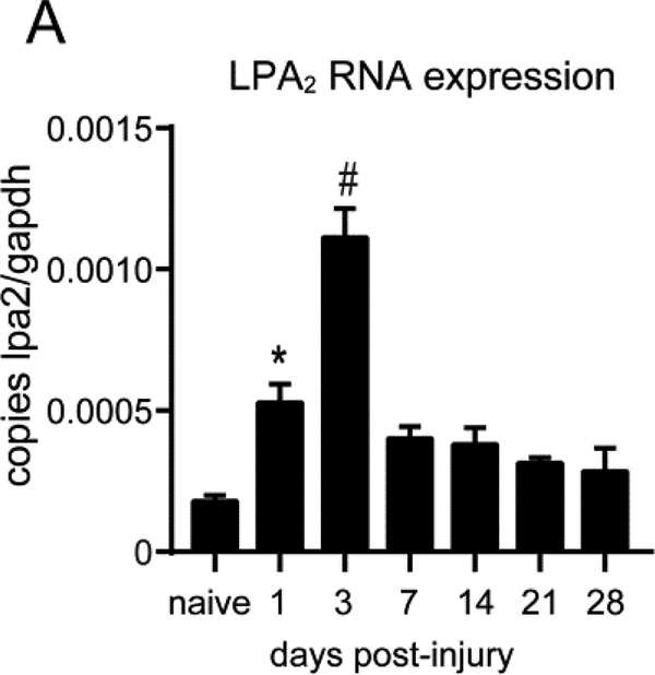
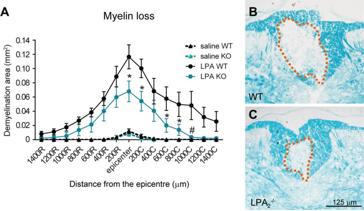
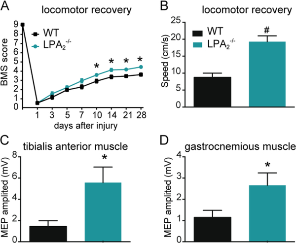
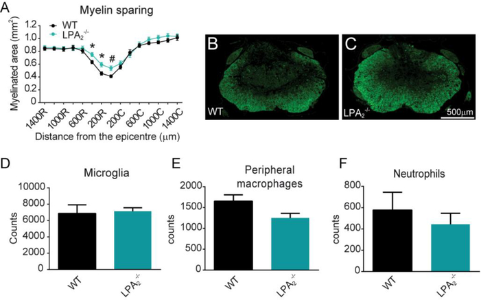
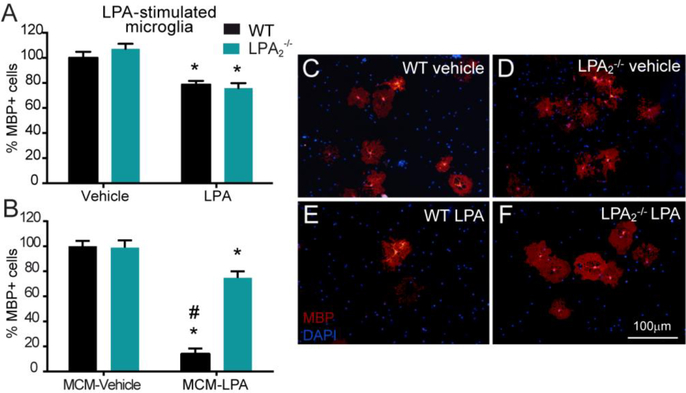
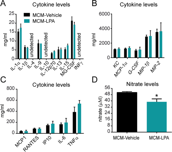
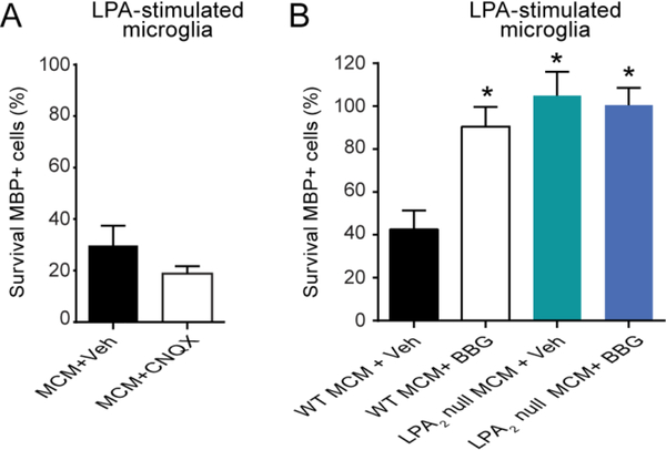
Lysophosphatidic acid (LPA) is an extracellular lipid mediator involved in many physiological functions by signaling through six known G-protein-coupled receptors (LPA1-LPA6). In the central nervous system (CNS), LPA mediates a wide range of effects, including neural progenitor cell physiology, astrocyte and microglia activation, neuronal cell death, axonal retraction, and contributions to pain, schizophrenia and hydrocephalus. We recently reported that LPA-LPA1 signaling mediates functional deficits and myelin loss after spinal cord injury (SCI). Here, we provide clear evidence on the deleterious contribution of another LPA receptor, LPA2, to myelin loss after SCI. We found that LPA2 is constitutively expressed in the spinal cord parenchyma and its transcripts were up-regulated after contusion injury, in part, by microglial cells. We also found that the demyelinating lesion triggered by intraspinal injection of LPA into the undamaged spinal cord was markedly reduced in the lack of LPA2. Similarly, LPA2 deficient mice showed enhanced motor skills and myelin sparing after SCI. To gain insights into the detrimental actions of LPA2 in spinal cord we performed cell culture studies. These experiments revealed that, similar to LPA1, activation of microglia LPA2 led to oligodendrocyte cell death. Moreover, we also found that the cytotoxic effects underlaying microglial LPA-LPA2 axis were mediated by the release of purines by microglia and the activation of P2X7 receptor on oligodendrocytes. Overall, this study provides new mechanistic insights into how LPA contributes to SCI physiopathology, and suggest that targeting LPA2 could be a novel therapeutic approach for the treatment of acute SCI.

Keywords: Demyelination; Inflammation; Lysophosphatidic acid; Microglia; Neurodegeneration; Spinal cord injury.


Figures

Figure 1.
Figure 1.. LPA2 mRNA expression is up-regulated in the injured spinal cord.
Real Time-PCR quantification of LPA2 mRNA levels from spinal cords harvested at different time points after contusion injury. (*p<0.05, #p<0.01; n=3 per time point). Error bars indicate SEM.
Figure 2.
Figure 2.. Lack of LPA2 reduces LPA-triggered demyelination.
(A) Quantification of myelin loss at various distances rostral and caudal to the injury epicenter. (B, C) Representative micrographs showing myelin loss at the injury epicenter in WT (B) and LPA2 deficient (C) mice spinal cords injected with 5 nmoles of LPA. (*p<0.05, #p<0.01; n=4 WT + saline, n=4 LPA2 null + saline, n=8 WT + LPA, n=7 LPA2 null + LPA). Error bars indicate SEM. Scale bar=125 μm.
Figure 3.
Figure 3.. Lack of LPA2 promotes functional recovery after SCI.
(A) Locomotor recovery after spinal cord contusion injury in WT and LPA2 null mice using the 9-point Basso Mouse Scale. The lack of LPA2 led to significant improvement in locomotor performance compared to WT mice (*p<0.05; n=12 for WT and 11 for LPA2 deficient mice). (B) The ability to run at a constant speed was evaluated at 28 dpi by placing mice onto the belt of a motorized treadmill. Note that mice lacking LPA2 were able to run at faster speeds. (#p<0.01; n=8 per group). (C, D) Preservation of descending axonal tracts after SCI was evaluated by registering the motor evoked potentials (MEPs) from tibialis anterior and gastrocnemius muscles. Mice lacking LPA2 showed higher MEP amplitudes for both muscles. (*p<0.05; n=12 for WT and 11 for LPA2 deficient mice). Error bars indicate SEM.
Figure 4.
Figure 4.. The lack of LPA2 results in reduced secondary damage after SCI.
(A) Quantification of myelin sparing at various distances rostral and caudal to the injury epicenter. (B, C) Representative micrographs of spinal cord tissue at the injury showing myelin sparing at the injury epicenter in WT (B) and LPA2 deficient (C) mice. (*p<0.05, #p<0.01 vs WT; n=12 for WT and 11 for LPA2 deficient mice). Error bars indicate SEM. Scale bar=500 μm. (D-E) Quantification of microglia, peripheral macrophages and neutrophils in the contused spinal cord of WT and LPA2 deficient mice at day 7 post-injury. (n=4 per group)
Figure 5.
Figure 5.. LPA induced oligodendrocyte cell death in vitro through LPA2 signaling on microglial cells.
(A) Quantification of MBP+ cells on primary cultured WT and LPA2 null oligodendrocytes exposed to 1 μM LPA for 24h. (*p<0.05; n=8 biological replicates for vehicle and n=7 biological replicates for LPA in both groups). (B) Quantification of MBP+ cells on primary cultured WT oligodendrocytes exposed for 24h to LPA-stimulated microglial-conditioned medium (MCM) from WT or LPA2 deficient mice. (*p<0.05, #p< 0.01 vs. control; n=8 biological replicates per group). (C-F) Representative micrographs showing MBP+ cells exposed to WT (C, E) or LPA2 null (D, F) MCM in control (C, D) or LPA (E, F) conditions. Error bars indicate SEM. Scale bar=100 μm
Figure 6.
Figure 6.. Cytokine and nitrate levels in LPA-stimulated microglia conditioned medium.
Measurement of protein levels for various cytokines. (n=4 biological replicates per group). (A, B, C) as well as nitrates (D) in the supernatant LPA-stimulated microglia conditioned medium (*p<0.05; n=3 per group). Error bars indicate SEM.
Figure 7.
Figure 7.. Effects of CNQX and BGG in oligodendrocyte survival after incubation with LPA-stimulated microglial conditioned medium.
(A) Quantification of MBP+ cells in oligodendrocyte cell cultures incubated with LPA-stimulated microglial conditioned medium. Note ~35% of oligodendrocyte survived after incubation with the conditioned medium of LPA-activated microglia, and that treatment of oligodendrocytes with the AMPA/Kainate receptor antagonist CNQX did not rescue cell death. (n=7 biological replicates per group). (B) Assessment of MBP+ cells after incubation of oligodendrocytes with LPA-stimulated WT or LPA2 null microglial conditioned in the presence of BBG. This P2X7 antagonist prevented oligodendrocyte death induced by LPA-stimulated WT microglia to similar levels to that triggered by the conditioned medium of LPA2 null microglia stimulated with LPA. However, BBG did not show additive effects on the LPA-stimulated LPA2 microglial conditioned medium. (*p<0.05 vs. Control; n=4 biological replicates per group). Error bars indicate SEM.

References

    1. Amo-Aparicio J, Martinez-Muriana A, Sanchez-Fernandez A, Lopez-Vales R, 2018. Neuroinflammation quantification for spinal cord injury. Curr. Protoc. Immunol 123:e57. - PubMed
    1. An S, Bleu T, Hallmark OG, Goetzl EJ, 1998. Characterization of a novel subtype of human G protein-coupled receptor for lysophosphatidic acid. J. Biol. Chem 273, 7906–7910. - PubMed
    1. Basso DM, Fisher LC, Anderson AJ, Jakeman LB, McTigue DM, Popovich PG, 2006. Basso Mouse Scale for locomotion detects differences in recovery after spinal cord injury in five common mouse strains. J. Neurotrauma 23, 635–659. - PubMed
    1. Brosius Lutz A, Barres BA, 2014. Contrasting the glial response to axon injury in the central and peripheral nervous systems. Dev. Cell 28, 7–17. - PubMed
    1. Choi JW, Chun J, 2013. Lysophospholipids and their receptors in the central nervous system. Biochimica et biophysica acta 1831, 20–32. - PMC - PubMed

Publication types

Substances