Transcription factor TFAP2B up-regulates human corneal endothelial cell-specific genes during corneal development and maintenance
- PMID: 30552118
- PMCID: PMC6378988
- DOI: 10.1074/jbc.RA118.005527
Transcription factor TFAP2B up-regulates human corneal endothelial cell-specific genes during corneal development and maintenance
Abstract
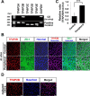
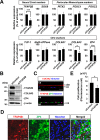
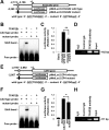
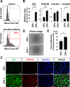
The corneal endothelium, which originates from the neural crest via the periocular mesenchyme (PM), is crucial for maintaining corneal transparency. The development of corneal endothelial cells (CECs) from the neural crest is accompanied by the expression of several transcription factors, but the contribution of some of these transcriptional regulators to CEC development is incompletely understood. Here, we focused on activating enhancer-binding protein 2 (TFAP2, AP-2), a neural crest-expressed transcription factor. Using semiquantitative/quantitative RT-PCR and reporter gene and biochemical assays, we found that, within the AP-2 family, the TFAP2B gene is the only one expressed in human CECs in vivo and that its expression is strongly localized to the peripheral region of the corneal endothelium. Furthermore, the TFAP2B protein was expressed both in vivo and in cultured CECs. During mouse development, TFAP2B expression began in the PM at embryonic day 11.5 and then in CECs during adulthood. siRNA-mediated knockdown of TFAP2B in CECs decreased the expression of the corneal endothelium-specific proteins type VIII collagen α2 (COL8A2) and zona pellucida glycoprotein 4 (ZP4) and suppressed cell proliferation. Of note, we also found that TFAP2B binds to the promoter of the COL8A2 and ZP4 genes. Furthermore, CECs that highly expressed ZP4 also highly expressed both TFAP2B and COL8A2 and showed high cell proliferation. These findings suggest that TFAP2B transcriptionally regulates CEC-specific genes and therefore may be an important transcriptional regulator of corneal endothelial development and homeostasis.
Keywords: COL8A2; TFAP2B; ZP4; cell surface protein; collagen; cornea; corneal endothelial cells; development; differentiation; eye; eye development; gene regulation; molecular cell biology; neural crest; regenerative medicine; transcriptional regulation.
© 2019 Hara et al.
Conflict of interest statement
The authors declare that they have no conflicts of interest with the contents of this article
Figures


Similar articles
-
AP-2β Is a Downstream Effector of PITX2 Required to Specify Endothelium and Establish Angiogenic Privilege During Corneal Development.Invest Ophthalmol Vis Sci. 2016 Mar;57(3):1072-81. doi: 10.1167/iovs.15-18103. Invest Ophthalmol Vis Sci. 2016. PMID: 26968737 Free PMC article.
-
Discovery of molecular markers to discriminate corneal endothelial cells in the human body.PLoS One. 2015 Mar 25;10(3):e0117581. doi: 10.1371/journal.pone.0117581. eCollection 2015. PLoS One. 2015. PMID: 25807145 Free PMC article.
-
Identification of Prdm genes in human corneal endothelium.Exp Eye Res. 2017 Jun;159:114-122. doi: 10.1016/j.exer.2017.02.009. Epub 2017 Feb 20. Exp Eye Res. 2017. PMID: 28228349 Free PMC article.
-
Challenges in corneal endothelial cell culture.Regen Med. 2021 Sep;16(9):871-891. doi: 10.2217/rme-2020-0202. Epub 2021 Aug 12. Regen Med. 2021. PMID: 34380324 Free PMC article. Review.
-
Towards Clinical Application: Calcium Waves for In Vitro Qualitative Assessment of Propagated Primary Human Corneal Endothelial Cells.Cells. 2024 Dec 5;13(23):2012. doi: 10.3390/cells13232012. Cells. 2024. PMID: 39682760 Free PMC article. Review.
Cited by
-
The Use of Induced Pluripotent Stem Cells as a Model for Developmental Eye Disorders.Front Cell Neurosci. 2020 Aug 20;14:265. doi: 10.3389/fncel.2020.00265. eCollection 2020. Front Cell Neurosci. 2020. PMID: 32973457 Free PMC article.
-
Revisiting Existing Evidence of Corneal Endothelial Progenitors and Their Potential Therapeutic Applications in Corneal Endothelial Dysfunction.Adv Ther. 2020 Mar;37(3):1034-1048. doi: 10.1007/s12325-020-01237-w. Epub 2020 Jan 30. Adv Ther. 2020. PMID: 32002810 Review.
-
TFAP2B overexpression contributes to tumor growth and progression of thyroid cancer through the COX-2 signaling pathway.Cell Death Dis. 2019 May 21;10(6):397. doi: 10.1038/s41419-019-1600-7. Cell Death Dis. 2019. PMID: 31113934 Free PMC article.
-
COL8A2 Regulates the Fate of Corneal Endothelial Cells.Invest Ophthalmol Vis Sci. 2020 Sep 1;61(11):26. doi: 10.1167/iovs.61.11.26. Invest Ophthalmol Vis Sci. 2020. PMID: 32931574 Free PMC article.
-
Directed Differentiation of Human Pluripotent Stem Cells towards Corneal Endothelial-Like Cells under Defined Conditions.Cells. 2021 Feb 5;10(2):331. doi: 10.3390/cells10020331. Cells. 2021. PMID: 33562615 Free PMC article.
References
Publication types
MeSH terms
Substances
LinkOut - more resources
Full Text Sources
Other Literature Sources
Research Materials

