Case Reports
. 2018 Dec 17;4(6):a003194.
doi: 10.1101/mcs.a003194. Print 2018 Dec.

A recurrent novel MGA-NUTM1 fusion identifies a new subtype of high-grade spindle cell sarcoma

Affiliations
Case Reports

A recurrent novel MGA-NUTM1 fusion identifies a new subtype of high-grade spindle cell sarcoma

Daniel Diolaiti et al. Cold Spring Harb Mol Case Stud. .

Abstract

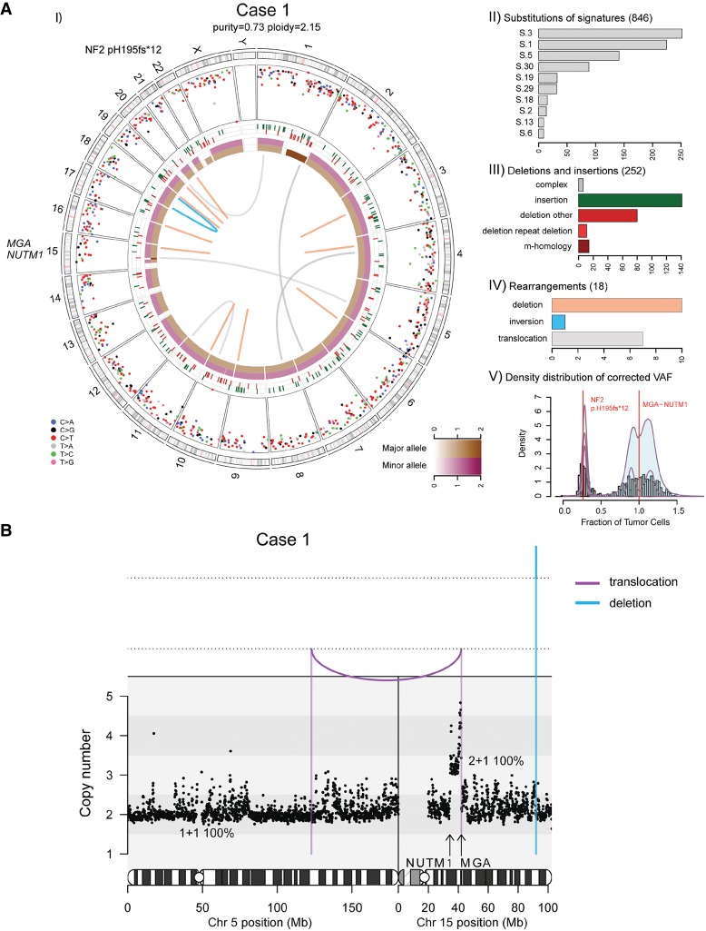
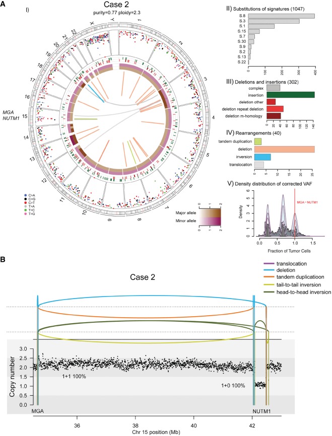
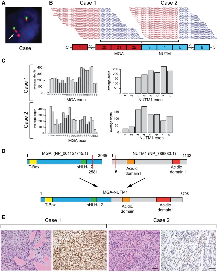
NUTM1-rearranged tumors are defined by the presence of a gene fusion between NUTM1 and various gene partners and typically follow a clinically aggressive disease course with poor outcomes despite conventional multimodality therapy. NUTM1-rearranged tumors display histologic features of a poorly differentiated carcinoma with areas of focal squamous differentiation and typically express the BRD4-NUTM1 fusion gene defining a distinct clinicopathologic entity-NUT carcinoma (NC). NCs with mesenchymal differentiation have rarely been described in the literature. In this report, we describe the characterization of two cases of high-grade spindle cell sarcoma harboring a novel MGA-NUTM1 fusion. Whole-genome sequencing identified the presence of complex rearrangements resulting in a MGA-NUTM1 fusion gene in the absence of other significant somatic mutations. Genetic rearrangement was confirmed by fluorescence in situ hybridization, and expression of the fusion gene product was confirmed by transcriptomic analysis. The fusion protein was predicted to retain nearly the entire protein sequence of both MGA (exons 1-22) and NUTM1 (exons 3-8). Histopathologically, both cases were high-grade spindle cell sarcomas without specific differentiation markers. In contrast to typical cases of NC, these cases were successfully treated with aggressive local control measures (surgery and radiation) and both patients remain alive without disease. These cases describe a new subtype of NUTM1-rearranged tumors warranting expansion of diagnostic testing to evaluate for the presence of MGA-NUTM1 or alternative NUTM1 gene fusions in the diagnostic workup of high-grade spindle cell sarcomas or small round blue cell tumors of ambiguous lineage.

Trial registration: ClinicalTrials.gov NCT01775072.

Keywords: MGA-NUT1; NUT carcinoma; NUTM1-rearranged tumor; spindle cell carcinoma; synovial sarcoma.


Figures

Figure 1.
Figure 1.
(A) Whole-genome analysis identifies a novel MGA–NUTM1 fusion in Case 1. (I) Circos plot summarizing the whole-genome sequencing (WGS) data. The two innermost tracks depict the integer copy-number changes for the major (brown) and minor (dark pink) allele. The outermost track shows the intermutation distance for substitutions each plotted according to the type of nucleotide change. The middle track shows the genomic positions of the small insertions (green) and deletions (red) along the genome. Rearrangements are plotted as arcs inside the Circos plot. Genes affected by potential oncogenic changes are annotated. (II) Mutation signature analysis of the substitutions using Mutational Patterns. Only the signatures with the 10 highest exposures are shown. (III) Summary of the indel data also showing the contribution of repeat- or microhomology-mediated mechanisms among deletions. (IV) Summary of rearrangement data. (V) Statistical analysis of the corrected variant allele frequency of substitution by Bayesian Dirichlet process–based clustering. Empiric histogram of substitutions is shown in gray together with the density from clustering in pale green and the fitted distribution in dark pink. Potential oncogenic alterations are annotated. (B) Integrated copy-number/rearrangement plots showing the WGS-based absolute copy number (y-axis) across the indicated genomic region (x-axis). Rearrangements are depicted as lines perpendicular to the x-axis.
Figure 2.
Figure 2.
(A) Whole-genome analysis identifies a novel MGA–NUTM1 fusion in Case 2. (I) Circos plot summarizing the whole-genome sequencing (WGS) data. (II) Mutation signature analysis of the substitutions using Mutational Patterns. (III) Summary of the indel data also showing the contribution of repeat- or microhomology-mediated mechanisms among deletions. (IV) Summary of rearrangement data. (V) Statistical analysis of the corrected variant allele frequency of substitution by Bayesian Dirichlet process–based clustering. For details, see legend to Figure 1. (B) Integrated copy-number/rearrangement plots showing the WGS-based absolute copy number (y-axis) across the indicated genomic region (x-axis). Rearrangements are depicted as lines perpendicular to the x-axis.
Figure 3.
Figure 3.
(A) Case 1: Fluorescence in situ hybridization assay showing evidence of complex NUTM1 rearrangements. The arrow indicates examples of abnormal doublet signals of the 3′ NUTM1 locus (orange). (B) RNA-sequencing reads spanning the junction between MGA exon 22 and NUTM1 exon 3. (C) RNA-seq coverage of MGA and NUTM1 exons. (D) Schematic representation of MGA and NUTM1 protein domains and resulting MGA–NUTM1 fusion protein structure. (E) H&E staining of both cases showed a monomorphic spindle cell morphology arranged in fascicles with an associated collagenous stroma. Case 1 showed dense hyalinized material, the so-called “amianthoid fibers,” resembling osteoid matrix deposition. Immunohistochemically Case 1 showed strong, diffuse nuclear reactivity for NUTM1, whereas Case 2 showed a multifocal weak staining pattern. Scale bars, 500 µM.

References

    1. Alekseyenko AA, Walsh EM, Zee BM, Pakozdi T, Hsi P, Lemieux ME, Dal Cin P, Ince TA, Kharchenko PV, Kuroda MI, et al. 2017. Ectopic protein interactions within BRD4-chromatin complexes drive oncogenic megadomain formation in NUT midline carcinoma. Proc Natl Acad Sci 114: E4184–E4192. - PMC - PubMed
    1. Alexandrov LB, Nik-Zainal S, Wedge DC, Aparicio SA, Behjati S, Biankin AV, Bignell GR, Bolli N, Borg A, Børresen-Dale AL, et al. 2013. Signatures of mutational processes in human cancer. Nature 500: 415–421. - PMC - PubMed
    1. Bauer DE, Mitchell CM, Strait KM, Lathan CS, Stelow EB, Lüer SC, Muhammed S, Evans AG, Sholl LM, Rosai J, et al. 2012. Clinicopathologic features and long-term outcomes of NUT midline carcinoma. Clin Cancer Res 18: 5773–5779. - PMC - PubMed
    1. Bolli N, Avet-Loiseau H, Wedge DC, Van Loo P, Alexandrov LB, Martincorena I, Dawson KJ, Iorio F, Nik-Zainal S, Bignell GR, et al. 2014. Heterogeneity of genomic evolution and mutational profiles in multiple myeloma. Nat Commun 5: 2997. - PMC - PubMed
    1. Burn SF, Washkowitz AJ, Gavrilov S, Papaioannou VE. 2018. Postimplantation Mga expression and embryonic lethality of two gene-trap alleles. Gene Expr Patterns 27: 31–35. - PMC - PubMed

Publication types

MeSH terms

Associated data