

Phototaxis in a wild isolate of the cyanobacterium Synechococcus elongatus
- PMID: 30552139
- PMCID: PMC6310787
- DOI: 10.1073/pnas.1812871115
Phototaxis in a wild isolate of the cyanobacterium Synechococcus elongatus
Abstract
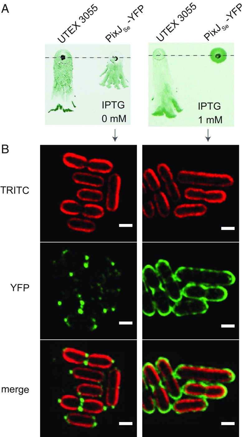
Many cyanobacteria, which use light as an energy source via photosynthesis, have evolved the ability to guide their movement toward or away from a light source. This process, termed "phototaxis," enables organisms to localize in optimal light environments for improved growth and fitness. Mechanisms of phototaxis have been studied in the coccoid cyanobacterium Synechocystis sp. strain PCC 6803, but the rod-shaped Synechococcus elongatus PCC 7942, studied for circadian rhythms and metabolic engineering, has no phototactic motility. In this study we report a recent environmental isolate of S. elongatus, the strain UTEX 3055, whose genome is 98.5% identical to that of PCC 7942 but which is motile and phototactic. A six-gene operon encoding chemotaxis-like proteins was confirmed to be involved in phototaxis. Environmental light signals are perceived by a cyanobacteriochrome, PixJSe (Synpcc7942_0858), which carries five GAF domains that are responsive to blue/green light and resemble those of PixJ from Synechocystis Plate-based phototaxis assays indicate that UTEX 3055 uses PixJSe to sense blue and green light. Mutation of conserved functional cysteine residues in different GAF domains indicates that PixJSe controls both positive and negative phototaxis, in contrast to the multiple proteins that are employed for implementing bidirectional phototaxis in Synechocystis.
Keywords: GAF domain; Synechococcus elongatus; cyanobacteria; photoreceptor; phototaxis.
Conflict of interest statement
The authors declare no conflict of interest.
Figures


Similar articles
-
Phototactic signaling network in rod-shaped cyanobacteria: A study on Synechococcus elongatus UTEX 3055.Microbiol Res. 2025 Mar;292:127967. doi: 10.1016/j.micres.2024.127967. Epub 2024 Nov 28. Microbiol Res. 2025. PMID: 39637757
-
The (PATAN)-CheY-Like Response Regulator PixE Interacts with the Motor ATPase PilB1 to Control Negative Phototaxis in the Cyanobacterium Synechocystis sp. PCC 6803.Plant Cell Physiol. 2020 Feb 1;61(2):296-307. doi: 10.1093/pcp/pcz194. Plant Cell Physiol. 2020. PMID: 31621869
-
Bipolar localization of putative photoreceptor protein for phototaxis in thermophilic cyanobacterium Synechococcus elongatus.Plant Cell Physiol. 2002 Dec;43(12):1585-8. doi: 10.1093/pcp/pcf176. Plant Cell Physiol. 2002. PMID: 12514256
-
Photosystems and photoreceptors in cyanobacterial phototaxis and photophobotaxis.FEBS Lett. 2024 Aug;598(15):1899-1908. doi: 10.1002/1873-3468.14968. Epub 2024 Jun 30. FEBS Lett. 2024. PMID: 38946046 Review.
-
Phototactic motility in the unicellular cyanobacterium Synechocystis sp. PCC 6803.Photochem Photobiol Sci. 2004 Jun;3(6):512-8. doi: 10.1039/b402320j. Epub 2004 May 11. Photochem Photobiol Sci. 2004. PMID: 15170479 Review.
Cited by
-
Recent developments in the production and utilization of photosynthetic microorganisms for food applications.Heliyon. 2023 Mar 22;9(4):e14708. doi: 10.1016/j.heliyon.2023.e14708. eCollection 2023 Apr. Heliyon. 2023. PMID: 37151658 Free PMC article. Review.
-
EbsA is essential for both motility and biofilm formation in the filamentous cyanobacterium Nostoc punctiforme.Microbiology (Reading). 2024 Sep;170(9):001498. doi: 10.1099/mic.0.001498. Microbiology (Reading). 2024. PMID: 39287971 Free PMC article.
-
Differential Phototactic Behavior of Closely Related Cyanobacterial Isolates from Yellowstone Hot Spring Biofilms.Appl Environ Microbiol. 2022 May 24;88(10):e0019622. doi: 10.1128/aem.00196-22. Epub 2022 May 2. Appl Environ Microbiol. 2022. PMID: 35499327 Free PMC article.
-
Transcriptomic and Phenomic Investigations Reveal Elements in Biofilm Repression and Formation in the Cyanobacterium Synechococcus elongatus PCC 7942.Front Microbiol. 2022 Jun 23;13:899150. doi: 10.3389/fmicb.2022.899150. eCollection 2022. Front Microbiol. 2022. PMID: 35814646 Free PMC article.
-
Photo-bioconvection: towards light control of flows in active suspensions.Philos Trans A Math Phys Eng Sci. 2020 Sep 4;378(2179):20190523. doi: 10.1098/rsta.2019.0523. Epub 2020 Aug 3. Philos Trans A Math Phys Eng Sci. 2020. PMID: 32762429 Free PMC article. Review.
References
-
- Armitage JP, Hellingwerf KJ. Light-induced behavioral responses (;phototaxis’) in prokaryotes. Photosynth Res. 2003;76:145–155. - PubMed
-
- Bhaya D. Light matters: Phototaxis and signal transduction in unicellular cyanobacteria. Mol Microbiol. 2004;53:745–754. - PubMed
-
- Schuergers N, Mullineaux CW, Wilde A. Cyanobacteria in motion. Curr Opin Plant Biol. 2017;37:109–115. - PubMed
Publication types
MeSH terms
Substances
Associated data
- Actions
- Actions
- Actions
Grants and funding
LinkOut - more resources
Full Text Sources
Molecular Biology Databases
