. 2019 Feb 20;39(8):1334-1346.
doi: 10.1523/JNEUROSCI.0352-18.2018. Epub 2018 Dec 14.

Loss of Adult 5-HT1A Autoreceptors Results in a Paradoxical Anxiogenic Response to Antidepressant Treatment

Affiliations

Loss of Adult 5-HT1A Autoreceptors Results in a Paradoxical Anxiogenic Response to Antidepressant Treatment

Valérie Turcotte-Cardin et al. J Neurosci. .

Abstract

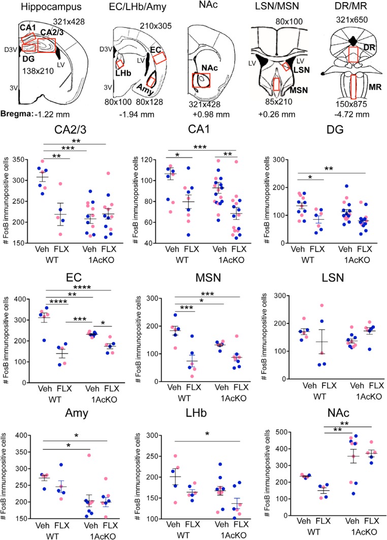
Selective serotonin (5-HT) reuptake inhibitors (SSRIs) are first-line antidepressants but require several weeks to elicit their actions. Chronic SSRI treatment induces desensitization of 5-HT1A autoreceptors to enhance 5-HT neurotransmission. Mice (both sexes) with gene deletion of 5-HT1A autoreceptors in adult 5-HT neurons (1AcKO) were tested for response to SSRIs. Tamoxifen-induced recombination in adult 1AcKO mice specifically reduced 5-HT1A autoreceptor levels. The 1AcKO mice showed a loss of 5-HT1A autoreceptor-mediated hypothermia and electrophysiological responses, but no changes in anxiety- or depression-like behavior. Subchronic fluoxetine (FLX) treatment induced an unexpected anxiogenic effect in 1AcKO mice in the novelty suppressed feeding and elevated plus maze tests, as did escitalopram in the novelty suppressed feeding test. No effect was seen in wild-type (WT) mice. Subchronic FLX increased 5-HT metabolism in prefrontal cortex, hippocampus, and raphe of 1AcKO but not WT mice, suggesting hyperactivation of 5-HT release. To detect chronic cellular activation, FosB+ cells were quantified. FosB+ cells were reduced in entorhinal cortex and hippocampus (CA2/3) and increased in dorsal raphe 5-HT cells of 1AcKO mice, suggesting increased raphe activation. In WT but not 1AcKO mice, FLX reduced FosB+ cells in the median raphe, hippocampus, entorhinal cortex, and median septum, which receive rich 5-HT projections. Thus, in the absence of 5-HT1A autoreceptors, SSRIs induce a paradoxical anxiogenic response. This may involve imbalance in activation of dorsal and median raphe to regulate septohippocampal or fimbria-fornix pathways. These results suggest that markedly reduced 5-HT1A autoreceptors may provide a marker for aberrant response to SSRI treatment.SIGNIFICANCE STATEMENT Serotonin-selective reuptake inhibitors (SSRIs) are effective in treating anxiety and depression in humans and mouse models. However, in some cases, SSRIs can increase anxiety, but the mechanisms involved are unclear. Here we show that, rather than enhancing SSRI benefits, adulthood knockout (KO) of the 5-HT1A autoreceptor, a critical negative regulator of 5-HT activity, results in an SSRI-induced anxiety effect that appears to involve a hyperactivation of the 5-HT system in certain brain areas. Thus, subjects with very low levels of 5-HT1A autoreceptors, such as during childhood or adolescence, may be at risk for an SSRI-induced anxiety response.

Keywords: 5-HT1A receptor; anxiety; autoreceptor; knock-out; raphe; serotonin.


Figures

Figure 1.
Figure 1.
Loss of 5-HT1A autoreceptors in adult 5-HT neurons of 1AcKO mice. A, Conditional KO strategy. To KO the HTR1A gene in adult 5-HT neurons, TPH2-CreERT2 and flx-5HT1A mice were bred to generate 1AcKO mice; upon treatment with tamoxifen, the 5-HT1A gene is knocked out and yellow fluorescent protein (YFP) is expressed in TPH+ serotonin neurons. B, C, Tamoxifen-induced YFP expression and 5-HT1A receptor loss in dorsal raphe (DR) 5-HT neurons. DR sections from tamoxifen-treated 1AcKO and WT littermates were stained for TPH (5-HT neuron marker) and YFP (marker of recombination) or 5-HT1A receptor. Scale bar is shown. D, E, Quantification of TPH/YFP- and TPH/5-HT1A-positive cells. Data are mean ± SE (n = 4), ****p < 0.0001 (unpaired two-tailed Student's t test). D, t = 152.9 df = 6; E, t = 90.39 df = 4. No YFP signal was detected in other brain regions (see Figure 1-1).
Figure 2.
Figure 2.
Loss of raphe 5-HT1A binding and autoreceptor function in 1AcKO mice. A, B, 5-HT1A receptor autoradiography of raphe and hippocampus. Top, Representative images of [125I]MPPI autoradiography of sections from 1AcKO and WT littermate mice showing dorsal (DR) and median (MR) raphe (A; boxes) and hippocampal (B; circumscribed) regions. C, D, Average intensity (μCi/region) was quantified for each region using templates above. Data are mean ± SEM (n = 3/group), unpaired two-tailed Student's t test. DR: **p = 0.0039, t = 16.05 df = 3; MR: *p = 0.0147, t = 5.088 df = 3; hippocampus: p = 0.1999, not significant, t = 1.440 df = 6. E, 5-HT1A-induced hypothermia. In WT, 8OH-DPAT (DPAT: 0.75 mg/kg, i.p.) induced body temperature reduction compared with saline (Sal), which was abolished in 1AcKO mice in both males and females. Data are mean ± SE (n = 7–12/group). Two-way ANOVA, Tukey's post-test showed significant changes for the following: WT-DPAT versus WT-Sal (black stars, below DPAT data); and WT-DPAT versus 1AcKO-DPAT (blue stars, between DPAT data); *p < 0.05; **p < 0.01; ***p < 0.001; ****p < 0.0001 (for statistics, see Figure 2-1; and Figure 2-2). The pretreatment temperatures (°C) were as follows (mean ± SD): M-SalWT, 38.0 ± 0.24; KO, 38.0 ± 0.24; M-DPAT WT, 38.6 ± 0.50; KO, 38.3 ± 0.31; F-SalWT, 38.1 ± 0.32; KO, 38.2 ± 0.47; F-DPAT WT, 38.2 ± 0.27; KO, 38.2 ± 0.31. F, Reduced 5-HT1A-mediated outward currents in 1AcKO mice. Whole-cell voltage-clamp recordings of DR 5-HT neurons from WT (n = 6) and 1AcKO (KO, n = 7) mice. Left, Time course of 5-HT1A-mediated outward current in response to 5-CT (100 nm; Vm = −55 mV). Right, Average peak steady-state 5-HT1AR-mediated currents from recorded 5-HT neurons. Data are mean ± SE. *p < 0.05 (unpaired two-tailed Student's t test).
Figure 3.
Figure 3.
Anxiogenic response to subchronic selective serotonin reuptake inhibitor (SSRI) treatment in 1AcKO mice. Top, Experimental timeline. At the start of antidepressant treatment, WT and 1AcKO mice were single-housed (SH), treated with tamoxifen (Tam), and treated with fluoxetine (FLX), escitalopram (ESC), or vehicle (Veh). After 9 d, the mice tested using the indicated behavior tests, maintaining treatment, and sacrificed at the end testing. Data points from individual male (M, blue) and female (F, pink) mice are shown. A, SSRI-induced anxiety in 1AcKO in the novelty suppressed feeding (NSF) test. Vehicle, FLX, and ESC were administered to WT (n (M/F (outlier)) = 9/8 (2), 8/11, 4/4, respectively) and 1AcKO (n = 6/4 (1), 5/7, 4/4, respectively) mice. Latency to feed in novel cage was recorded. FLX versus Veh: F(3,53) = 7.941, p = 0.0002; ESC versus Veh, F(3,38) = 3.172, p = 0.0351. B, FLX-induced anxiety in 1AcKO mice in the elevated plus maze (EPM) test. Vehicle, FLX, and ESC were administered to WT (n = 9/8, 8/11, 4/4, respectively) and KO (n = 6/6, 5/8 (1), 4/4, respectively), and time spent in the open arm was measured. FLX versus VEH: F(3,56) = 4.128, p = 0.0103; ESC versus VEH: F(3,40) = 1.938, p = 0.1389. C, D, Tail suspension (TS) and forced swim test (FST). For TS: WT, n (M/F (outlier)) = 3/3, 4/5, 4/4; KO, n = 2/3, 4/3, 4/4; for FST: WT n = 5/5, 7/6, 4/4; KO, n = 2/3 (1), 3/3, 4/4, for Veh, FLX, ESC, respectively. No effect of FLX or ESC was seen on immobility time in these two tests of depression-like behavior. TS versus VEH: FLX, F(3,23) = 0.6195, p = 0.6095; ESC, F(3,23) = 0.6724, p = 0.5777; FST: FLX, F(1,30) = 0.0358, p = 0.8512; ESC, F(1,27) = 1.458, p = 0.2377. Data are mean ± SE. *p < 0.05; **p < 0.01; ***p < 0.001 for drug versus VEH, or as indicated, by two-way ANOVA, Tukey's post-test. No differences between genotypes were observed in control measures for NSF (latency to feed, food consumption), EPM (closed arm time), or in the LD test (see Figure 3-1).
Figure 4.
Figure 4.
Enhanced 5-hydroxy indole acetic acid (5-HIAA)/5-HT in FLX-treated 1AcKO mice. Tissue 5-HT and 5-HIAA content in WT and 1AcKO mice was quantified in prefrontal cortex (PFC), hippocampus (Hippo), and dorsal raphe (DR) following fluoxetine (FLX) or vehicle (Veh) treatment (21–23 d), and the 5-HIAA/5-HT ratio calculated. Data points from individual male (M, blue) and female (F, pink) mice are shown. FLX treatment increased 5-HIAA/5-HT ratio in all 1AcKO tissues, and a significant reduction in 5-HT and increase in 5-HIAA was seen in FLX-treated 1AcKO PFC and hippocampus, whereas only partial or no effects were seen in WT. For WT (Veh, FLX), n (M/F) = 3/3, 3/3; for KO, n = 3/3, 5/5. Data are mean ± SE. *p < 0.05 (two-way ANOVA, Tukey's post-test). **p < 0.01 (two-way ANOVA, Tukey's post-test). ***p < 0.001 (two-way ANOVA, Tukey'spost-test). ****p < 0.0001 (two-way ANOVA, Tukey'spost-test). For treatment × genotype interaction, see Figure 4-1.
Figure 5.
Figure 5.
Opposite fluoxetine (FLX)-induced FosB changes in 5-HT neurons of 1AcKO versus WT mice. Vehicle (Veh) or FLX was administered to WT and 1AcKO mice for 24 d. A, Merge of immunofluorescence staining of dorsal (DR) and median (MR) raphe nuclei using DAPI (nuclei), anti-TPH (5-HT marker), and anti-FosB (chronic activity marker) from WT (left) or 1AcKO (right) mice. Carat symbols represent examples of FosB/TPH+ cells. For images of single staining, see Figure 5-2. B, C, Quantification of total TPH+, FosB+, and FosB/TPH+ cells in dorsal (B) and median (C) raphe for male (M, blue) and female (F, pink) mice. In dorsal raphe, FLX reduced TPH/FosB+ cells in 1AcKO but not WT; in median raphe, FLX reduced TPH/FosB+ cells in WT but not 1AcKO. For DR (TPH+, FosB+, TPH/FosB+), n (M/F (outliers)) = 3/4, 3/4, 4/4 (WT-Veh); 4/5, 5/4, 6/5 (WT-FLX); 5/6, 5/5, 8/8 (1) (KO-Veh); 7/5, 8/5, 11/9 (1) (KO-FLX); for MR, n = 5/5, 5/5, 5/5 (1) (WT-Veh); 4/4, 3/3, 4/5 (WT-FLX); 5/6, 5/6, 5/5 (1) (KO-Veh); 6/6, 5/6, 6/5 (1) (KO-FLX). Significant treatment × genotype interaction was detected for FosB/TPH+ cells in both DR and MR (Figure 5-1. Data are mean ± SE. *p < 0.05 (two-way ANOVA, Tukey's post-test). ***p < 0.001 (two-way ANOVA, Tukey's post-test).
Figure 6.
Figure 6.
Brain regional differences in FLX-induced FosB changes in 1AcKO versus WT mice. Shown above are figurative images of coronal sections at bregma levels (Paxinos and Franklin, 2001) and including regions of interest and templates (μm × μm) used for cell counting. Vehicle (Veh) or FLX was administered to mice for 24 d. Immunofluorescence staining for FosB was compared in 1AcKO and WT brain sections showing detectable FosB, and FosB+ cells were quantified and plotted for male (M, blue) and female (F, pink) mice. For WT-Veh, FLX and KO-Veh, FLX, n (M/F (outliers)): hippocampal CA2/3 (4/3, 3/2, 6/6 (1), 6/6 (2)), CA1 (4/4, 4/4, 11/8 (2), 7/8), DG (6/6, 4/3, 9/7 (1), 9/6 (2)); EC (3/3, 3/2, 4/3 (2), 3/3 (2)); MSN (3/3, 3/3, 4/4 (2), 4/4); LSN (3/3, 3/3, 5/5, 4/3); Amy (2/3 (1), 2/1 (2), 5/4 (1), 3/4); LHb (3/2 (1), 2/2 (1), 5/5 (1), 4/3 (1)); NAc (2/3, 2/3, 5/4 (1), 2/3 (2)). Significant treatment × genotype interaction was detected for EC, CA2/3, MSN, with trend for LSN (Figure 6-1). Data are mean ± SE. Two-way ANOVA, Tukey's post-test: *p < 0.05; **p < 0.01; ***p < 0.001; ****p < 0.0001.

Similar articles

Cited by

References

    1. aan het Rot M, Mathew SJ, Charney DS (2009) Neurobiological mechanisms in major depressive disorder. CMAJ 180:305–313. 10.1503/cmaj.080697 - DOI - PMC - PubMed
    1. Albert PR. (2012) Transcriptional regulation of the 5-HT1A receptor: implications for mental illness. Philos Trans R Soc Lond B Biol Sci 367:2402–2415. 10.1098/rstb.2011.0376 - DOI - PMC - PubMed
    1. Albert PR, Lemonde S (2004) 5-HT1A receptors, gene repression, and depression: guilt by association. Neuroscientist 10:575–593. 10.1177/1073858404267382 - DOI - PubMed
    1. Albert PR, Lembo P, Storring JM, Charest A, Saucier C (1996) The 5-HT1A receptor: signaling, desensitization, and gene transcription. Neuropsychopharmacology 14:19–25. 10.1016/S0893-133X(96)80055-8 - DOI - PubMed
    1. Albert PR, Vahid-Ansari F, Luckhart C (2014) Serotonin-prefrontal cortical circuitry in anxiety and depression phenotypes: pivotal role of pre- and post-synaptic 5-HT1A receptor expression. Front Behav Neurosci 8:199. 10.3389/fnbeh.2014.00199 - DOI - PMC - PubMed

Publication types

MeSH terms