. 2019 Mar;68(3):395-405.
doi: 10.1007/s00262-018-2280-3. Epub 2018 Dec 14.

Ex vivo conditioning with IL-12 protects tumor-infiltrating CD8+ T cells from negative regulation by local IFN-γ

Affiliations

Ex vivo conditioning with IL-12 protects tumor-infiltrating CD8+ T cells from negative regulation by local IFN-γ

Lin Lin et al. Cancer Immunol Immunother. 2019 Mar.

Abstract

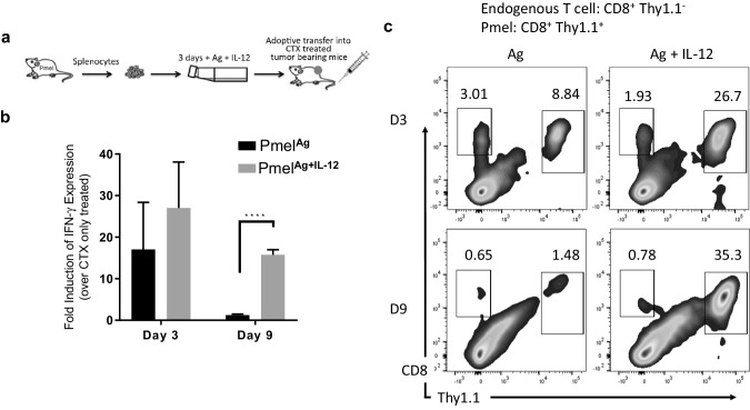
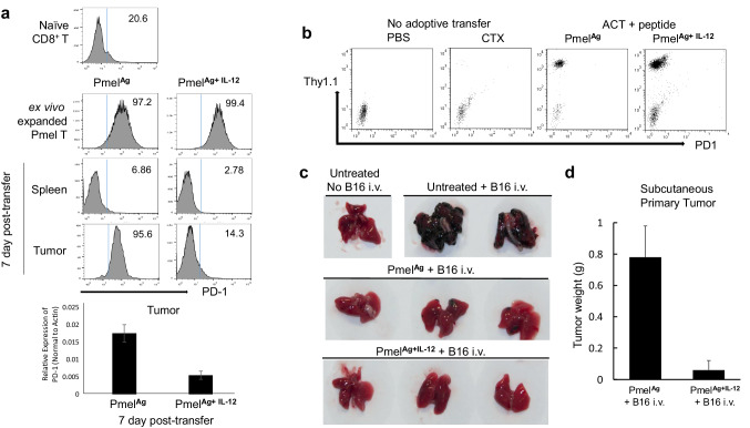
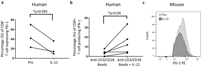
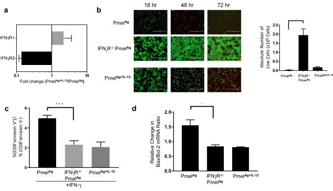
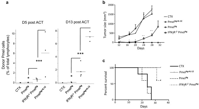
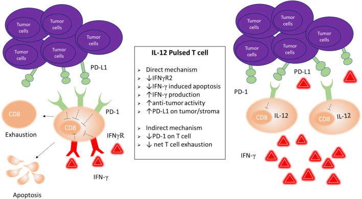
Optimal ex vivo expansion protocols for adoptive cell therapy (ACT) must yield T cells able to effectively home to tumors and survive the inhospitable conditions of the tumor microenvironment (TME), while simultaneously exerting persistent anti-tumor effector functions. Our previous work has shown that ex vivo activation in the presence of IL-12 can induce optimal expansion of murine CD8+ T cells, thus resulting in significant tumor regression after ACT mostly via sustained secretion of IFN-γ. In this report, we further elucidate the mechanism of this potency, showing that IL-12 additionally counteracts the negative regulatory effects of autocrine IFN-γ. IL-12 not only downregulates PD-1 expression by T cells, thus minimizing the effects of IFN-γ-induced PD-L1 upregulation by tumor stromal cells, but also inhibits IFNγR2 expression, thereby protecting T cells from IFN-γ-induced cell death. Thus, the enhanced anti-tumor activity of CD8+ T cells expanded ex vivo in the presence of IL-12 is due not only to the ability of IL-12-stimulated cells to secrete sustained levels of IFN-γ, but also to the additional capacity of IL-12 to counter the negative regulatory effects of autocrine IFN-γ.

Keywords: Adoptive T cells transfer; Melanoma/skin cancers; Models of host–tumor interactions; PD-1; Tumor microenvironment; Tumor promotion and progression.


Conflict of interest statement

The authors declare that they have no conflict of interest.

Figures

Fig. 1
Fig. 1
Anti-tumor activity of PmelAg+IL-12 cells associates with sustained levels of IFN-γ. a Adoptive cell therapy approach involving the infusion of Pmel cells activated in the presence of antigen with or without IL-12 into a lymphopenic host. This approach mediates the regression of pre-established melanoma lesions which results in improved survival. b Tumors from mice treated with PmelAg or PmelAg+IL-12 were harvested 3 and 9 days after adoptive transfer. Expression of IFN-γ in RNA isolated from tumors was measured using SYBR green quantitative real-time PCR. The relative quantification of the target transcripts normalized to the endogenous control (actin) was determined by the comparative Ct method. Data are presented as average fold change ± SD (n = 3) relative to tumors from control mice that did not receive adoptive transfer. Two-tailed Student’s t test was used, *p < 0.05, **p < 0.005, and ***p < 0.0001. c Tumors were digested to single cell suspensions and analyzed for frequency of infiltrating Pmel cells (Thy1.1+) by flow cytometry. Data shown are representative of at least three independent experiments
Fig. 2
Fig. 2
IL-12 downregulates PD-1 expression on adoptively transferred TILs independently of antigen. a Expression of PD-1 among Pmel cells activated in the presence of antigen plus or minus IL-12 prior to and 7 days after adoptive transfer in cell suspensions from spleen and tumor was determined by flow cytometry. Decreased levels of PD-1 expression among tumor-infiltrating PmelAg+IL-12 was confirmed by quantitative real-time PCR. b Mice bearing B16 melanoma tumors treated with PmelAg, PmelAg+IL-12, or CTX or PBS alone were injected i.p with 50 µg of cognate peptide every other day for 10 days. At day 11, blood was collected and expression of PD-1 was assessed on circulating Pmel (Thy1.1+) cells. c 10 days after transfer, mice bearing B16 melanoma tumors untreated or treated with PmelAg or PmelAg+IL-12 cells were injected i.v. with 5 × 104 B16 melanoma cells. 25 days after transfer mice were killed and lungs excised and examined for the presence of B16 nodules. d At the same time, primary tumors were excised and weighed. Tumor size is presented as the average weight ± SD (n = 3). Data shown are representative of at least three independent experiments
Fig. 3
Fig. 3
IL-12 can directly downregulate the expression of PD-1 on human TILs. a Tumor cell suspensions from human melanoma tumors were incubated with 10 ng/ml human IL-12 for 72 h, and the expression of PD-1 among CD8+ T cells was determined by flow cytometry before and after culture. (n = 3). b The same tumor cell suspensions were cultured in the presence of anti-CD3/CD28 beads with or without IL-12, and production of IFN-γ among CD8+ T cells was determined by flow cytometry (n = 5). One-tailed paired Student’s t test was used, *p < 0.05, **p < 0.005, and ***p < 0.0001. c Cell suspensions from a B16 melanoma tumor were incubated with 10 ng/ml murine IL-12 for 72 h, and the expression of PD-1 among CD8+ T cells was determined by flow cytometry
Fig. 4
Fig. 4
Lack of IFN-γ sensitivity protects activated CD8 + T cells from IFN-γ-induced apoptosis. a Adoptively transferred Thy1.1+ Pmel cells were sorted from cell suspensions prepared from similar size tumors (n = 3) 5 days after treatment with PmelAg or PmelAg+IL-12 cells, and analyzed for IFNγR1 and IFNγR2 expression by qPCR. Data are expressed as fold expression of PmelAg+IL-12 over PmelAg ±SD (p = 0.07). 1 × 106 ex vivo expanded wild-type PmelAg or PmelAg+IL-12, and IFNγR−/− PmelAg cell were recultured in fresh media. b Cell viability was determined after 24, 48, and 72 h of culture using the LIVE/DEAD® assay. c Apoptosis was measured by annexin V stain 24 h after culture. Data are presented as the average ratio of CD8+ annexin V+ over CD8+ annexin V cells ± SD (n = 3). d RNA isolated from cells after 24 h of culture was analyzed by qPCR for expression of Bcl-2 and Bax. These values were used to calculate Bax/Bcl-2 ratio which directly associates with apoptosis [20]. Data are presented as the average change in Bax/Bcl-2 ratio relative to their respective baseline measurement ± SD (n = 3). Two-tailed Student’s t test was used, *p < 0.05, **p < 0.005, and ***p < 0.0001. Data shown are representative of at least three independent experiments
Fig. 5
Fig. 5
Adoptively transferred ex vivo expanded Pmel cells unable to respond to IFN-γ stimuli have longer in vivo survival and superior anti-tumor activity. a Circulating levels of adoptively transferred PmelAg or IFNγR−/− PmelAg ex vivo expanded in the presence of antigen plus or minus IL-12 at days 5 and 13 after infusion into wild-type mice bearing B16 melanoma tumors. Data are presented as the average percent of CD8+/Thy1.1+ cells of total lymphocytes ± SD. (n = 4–5). b Tumor progression and c overall survival of mice treated with CTX alone or with either PmelAg, PmelAg+12 or IFNγR−/− PmelAg cells. Two-tailed Student’s t test was used, *p < 0.05, **p < 0.005, and ***p < 0.0001. Cumulative survival was calculated using a Kaplan–Meier curve using GraphPad Prism (n = 5). Data shown are representative of at least three independent experiments
Fig. 6
Fig. 6
IL-12-induced mechanisms to counter negative feedback by IFN-γ Exposure to IL-12 during ex vivo expansion protects tumor-infiltrating transferred CD8+ T cells from negative feedback by IFN-γ, directly by downregulating IFNγR2 which results in reduced apoptosis, and indirectly by downregulating PD-1 hence circumventing the impact of IFN-γ-induced PD-L1 upregulation on tumor stromal cells

References

    1. Rosenberg SA, Restifo NP, Yang JC, Morgan RA, Dudley ME. Adoptive cell transfer: a clinical path to effective cancer immunotherapy. Nat Rev Cancer. 2008;8(4):299–308. doi: 10.1038/nrc2355. - DOI - PMC - PubMed
    1. Curtsinger JM, Schmidt CS, Mondino A, Lins DC, Kedl RM, Jenkins MK, Mescher MF. Inflammatory cytokines provide a third signal for activation of naive CD4 + and CD8+ T cells. J Immunol. 1999;162(6):3256–3262. - PubMed
    1. Valenzuela J, Schmidt C, Mescher M. The roles of IL-12 in providing a third signal for clonal expansion of naive CD8 T cells. J Immunol. 2002;169(12):6842–6849. doi: 10.4049/jimmunol.169.12.6842. - DOI - PubMed
    1. Mescher MF, Curtsinger JM, Agarwal P, Casey KA, Gerner M, Hammerbeck CD, Popescu F, Xiao Z. Signals required for programming effector and memory development by CD8+ T cells. Immunol Rev. 2006;211:81–92. doi: 10.1111/j.0105-2896.2006.00382.x. - DOI - PubMed
    1. Chowdhury FZ, Ramos HJ, Davis LS, Forman J, Farrar JD. IL-12 selectively programs effector pathways that are stably expressed in human CD8+ effector memory T cells in vivo. Blood. 2011;118(14):3890–3900. doi: 10.1182/blood-2011-05-357111. - DOI - PMC - PubMed

MeSH terms