Review
. 2020 Jun;23(2):127-137.
doi: 10.1007/s40477-018-0349-7. Epub 2018 Dec 14.

Pediatric encephalic ultrasonography: the essentials

Affiliations
Review

Pediatric encephalic ultrasonography: the essentials

Valerio Vitale et al. J Ultrasound. 2020 Jun.

Abstract

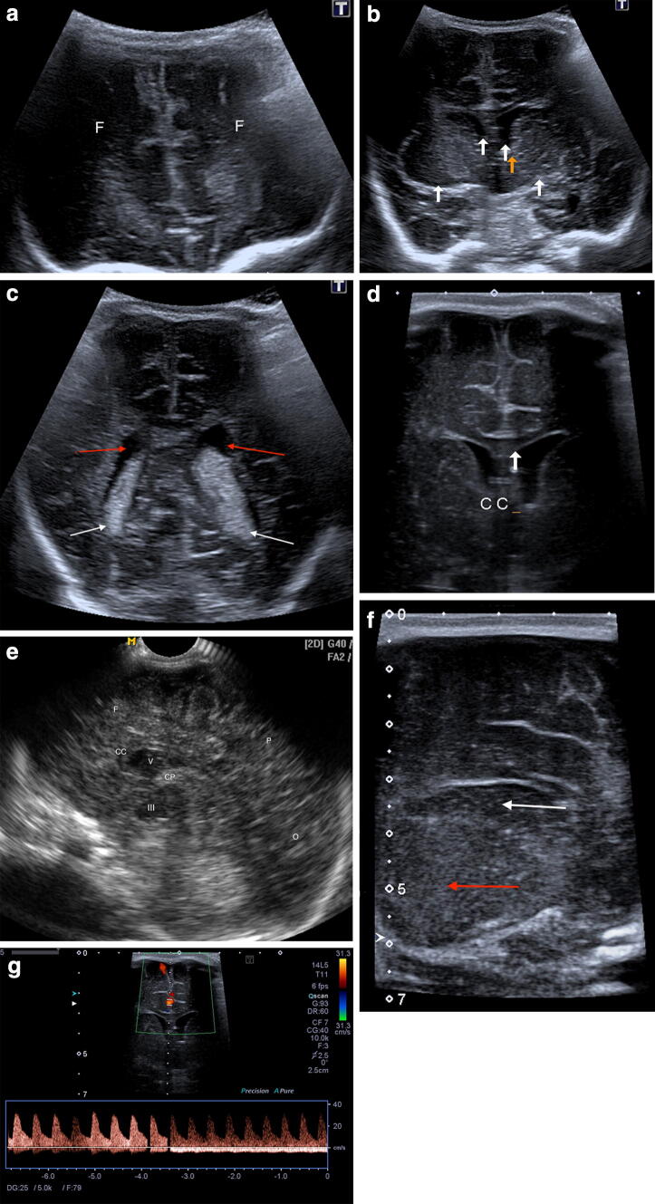
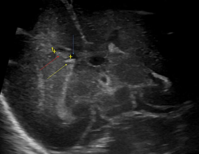
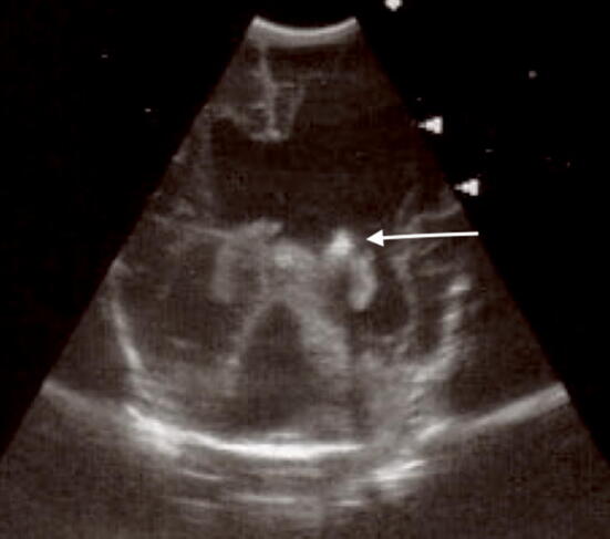
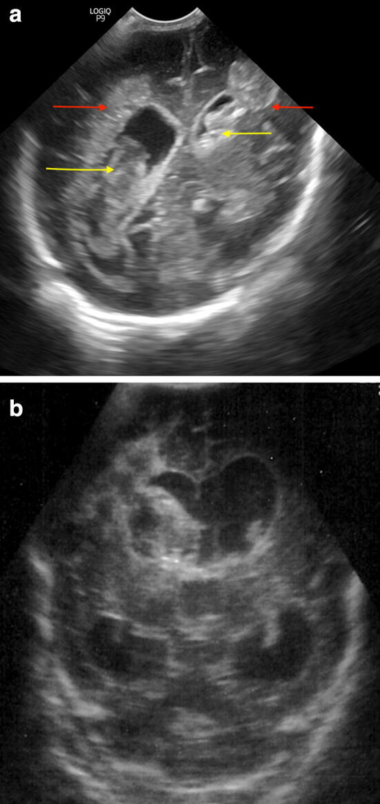
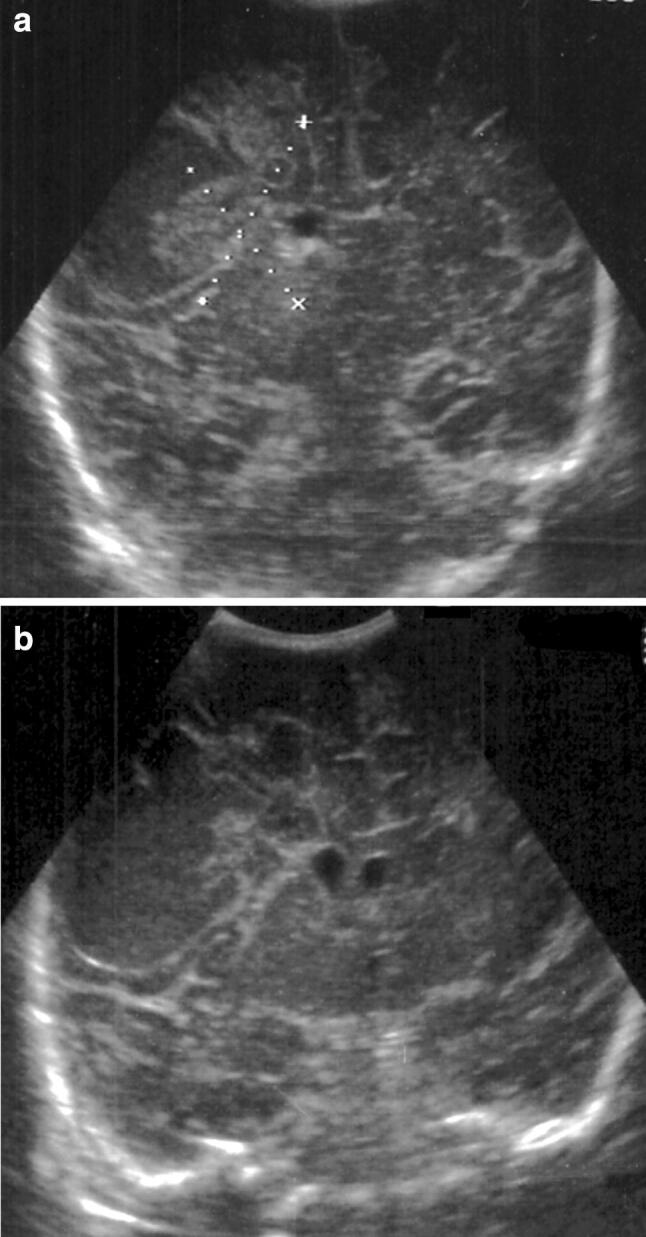
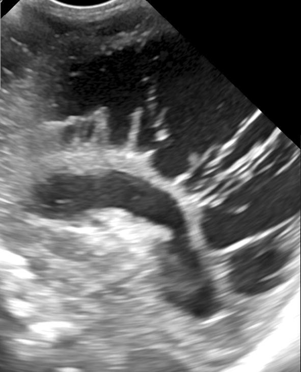
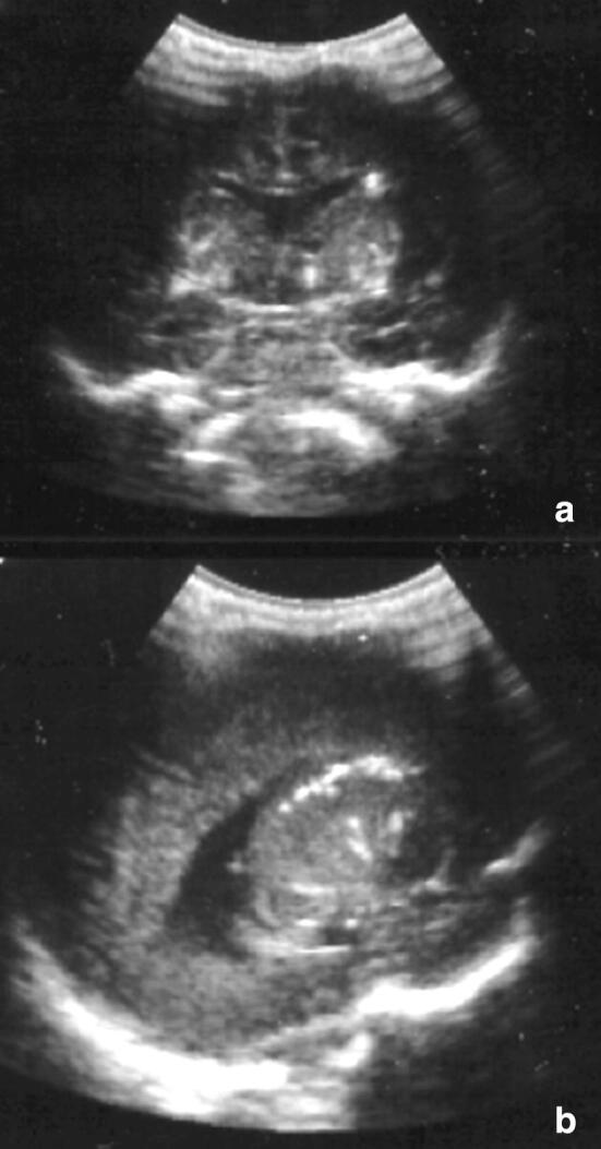
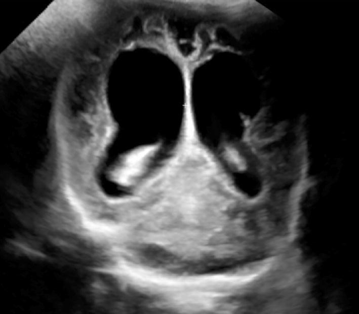
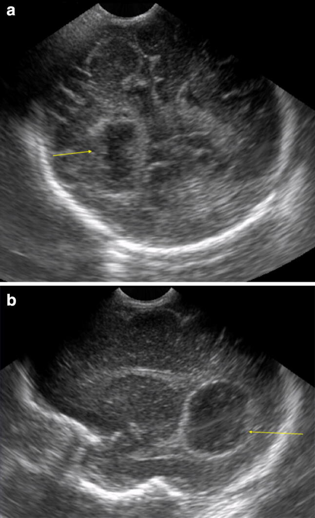
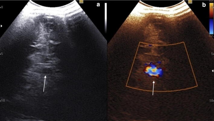
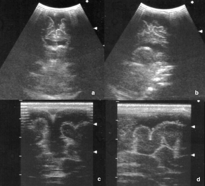
Nowadays, cranial ultrasonography (US) of the newborn represents the first imaging method in brain damage study and its possible outcomes. This exam is performed using the natural fontanelles, especially the anterior one. It is fast, non-invasive and does not produce any side effect. Ultrasonographic examination is usually performed in cases of prematurity, especially in children with birth weight less than 1500 g, because important informations about the possible presence of pathologies such as cerebral hemorrhage and hypoxic-ischemic encephalopathy are given. This approach can be useful also in the study of pre- and post-natal infections, for example, type II Herpes Simplex virus or Cytomegalovirus infections, or pointing out vascular malformations such as vein of Galen aneurysm. Although less important than methods such as computed tomography (CT) and magnetic resonance imaging (MRI) in the evaluation of trauma and tumors, ultrasound can provide useful informations or be used in first instance in the suspicion of a brain mass.

Al giorno d’oggi l’ecografia encefalica del neonato rappresenta la prima metodica di imaging nello studio del danno cerebrale e dei suoi possibili esiti. Tale esame viene eseguito utilizzando le fontanelle naturali, in particolare quella anteriore. È veloce, non invasivo e privo di complicanze. L’esame ecografico è di solito eseguito in caso di prematurità, soprattutto nei bambini con un peso alla nascita inferiore a 1500 grammi, potendo fornire importanti informazioni riguardo la presenza di patologie come l’emorragia cerebrale e l’encefalopatia ipossico-ischemica. Questo approccio è utile anche per lo studio delle infezioni pre- e post-natali, quali ad esempio quelle da virus Herpes Simplex di tipo II o da Citomegalovirus, o per evidenziare malformazioni vascolari quali l’aneurisma della vena di Galeno. Sebbene meno importante rispetto a metodiche quali la tomografia computerizzata (TC) e risonanza magnetica (RM) nello studio dei traumi e dei tumori, l’ecografia può talvolta fornire informazioni utili o essere la metodica di prima istanza nel sospetto di una massa cerebrale.

Keywords: Brain; Hemorrhage; Pediatrics; Periventricular leukomalacia; Ultrasonography.


Conflict of interest statement

The authors declare that they have no conflict of interest.

Figures

Fig. 1
Fig. 1
Normal anatomy. Coronal anterior (a), median (b superior white arrows lateral ventricles, inferior white arrows basal ganglia, orange arrow zone of caudothalamic notch) and posterior (c, white arrows choroid plexus, red arrow lateral ventricles) scan with magnified coronal scan (d) showing corpus callosum; g magnified coronal scan with normal color Doppler signal in cerebral anterior artery. Sagittal scan (e) and magnified parasagittal scan (f, thalamus red arrow and caudate nucleus white arrow). CC corpus callosum, III third ventricle, F frontal lobes, V lateral ventricle, O occipital lobe, P parietal lobe, CP choroid plexus
Fig. 2
Fig. 2
a Coronal scan. Hyperechoic subependymal hemorrhage (grade I, red arrow and calipers) without ventricular involvement (blue arrow thalamus; green arrow caudate nucleus), b coronal median scan with presence of a cystic area after 10 days from a grade I hemorrhage (white arrow)
Fig. 3
Fig. 3
Coronal scan. Hyperechoic right intraventricular hemorrhage (grade II, red arrow; yellow arrow choroid plexus; blue arrow normal lateral ventricle)
Fig. 4
Fig. 4
Coronal scan. Ventricular enlargement with the presence of derivation catheter (white arrow) after grade III hemorrhage
Fig. 5
Fig. 5
a Coronal scan. Intraparenchymal (red arrows) and intraventricular (yellow arrows) bilateral hemorrhage (grade IV); b Coronal scans showing right grade IV hemorrhage
Fig. 6
Fig. 6
Coronal scans. Right fronto-temporal subacute–chronic parenchymal hemorrhage with initial cystic malacic cavitation
Fig. 7
Fig. 7
Sagittal scan showing large hypoechoic areas of cystic PVL
Fig. 8
Fig. 8
Coronal (a) and sagittal (b) scans. Multiple small periventricular hyperechoic spots in Cytomegalovirus infection
Fig. 9
Fig. 9
Cerebral atrophy and ventricular enlargement after herpes simplex virus encephalitis
Fig. 10
Fig. 10
Coronal posterior (a) and sagittal (b) scans. Right abscessual round cavity (yellow arrow)
Fig. 11
Fig. 11
Coronal scans showing a posterior median pseudo-cystic lesion with internal vascular signal, Galen aneurysm malformation (white arrow)
Fig. 12
Fig. 12
a, b Coronal and sagittal scans showing a hypoechoic subdural fluid collection; c, d magnified view of the same patient

References

    1. Stanojevic M, Hafner T, Kurjak A. Three-dimensional (3D) ultrasound—a useful imaging technique in the assessment of neonatal brain. J Perinat Med. 2002;30(1):74–83. doi: 10.1515/JPM.2002.010. - DOI - PubMed
    1. Slovis TL, Kuhns LR. Real-time sonography of the brain through the anterior fontanelle. Am J Roentgenol. 1981;136:277–286. doi: 10.2214/ajr.136.2.277. - DOI - PubMed
    1. Di Salvo DN. A new view of the neonatal brain: clinical utility of supplemental neurologic US imaging windows. RadioGraphics. 2001;21:943–955. doi: 10.1148/radiographics.21.4.g01jl14943. - DOI - PubMed
    1. Bhat V, Bhat V. Neonatal neurosonography: a pictorial essay. Indian J Radiol Imaging. 2014;24(4):389–400. doi: 10.4103/0971-3026.143901. - DOI - PMC - PubMed
    1. Lowe LH, Bailey Z. State-of-the-art cranial sonography: part 1, modern techniques and image interpretation. Am J Roentgenol. 2011;196:1028–1033. doi: 10.2214/AJR.10.6160. - DOI - PubMed