. 2018 Dec 15;17(1):174.
doi: 10.1186/s12943-018-0919-5.

C-Src confers resistance to mitotic stress through inhibition DMAP1/Bub3 complex formation in pancreatic cancer

Affiliations

C-Src confers resistance to mitotic stress through inhibition DMAP1/Bub3 complex formation in pancreatic cancer

Jingjie Li et al. Mol Cancer. .

Erratum in

Abstract

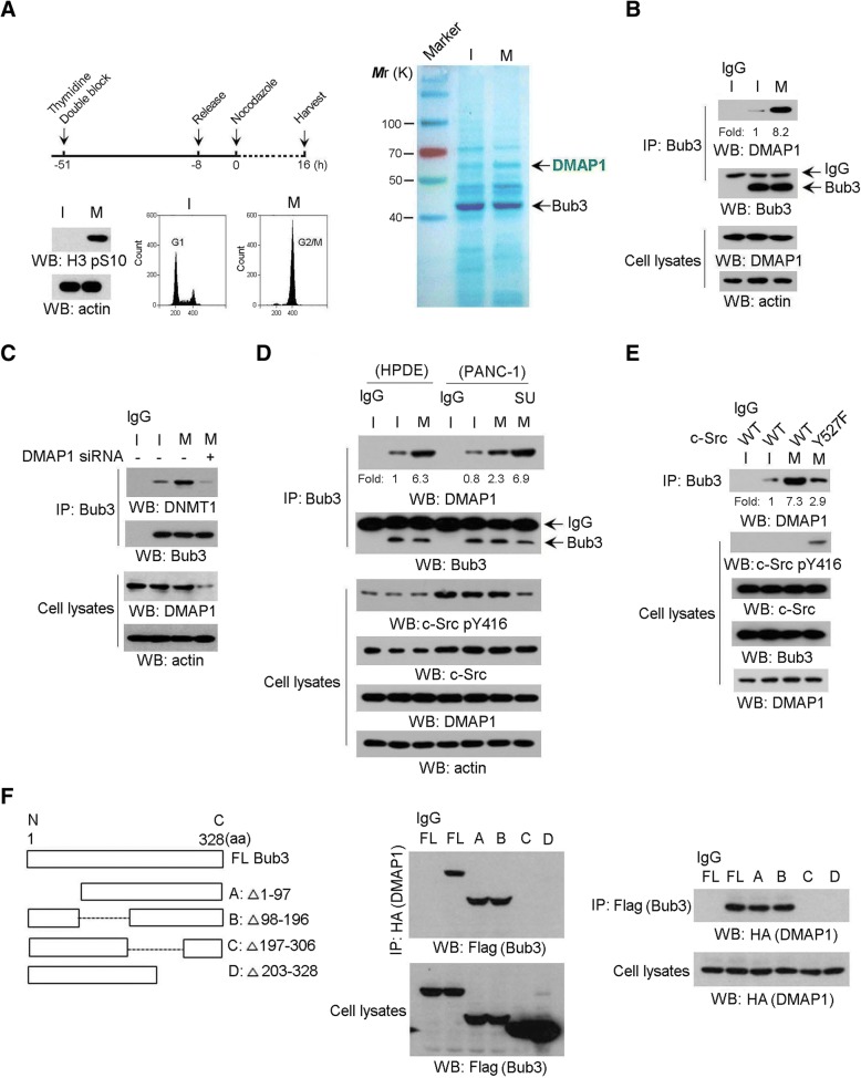
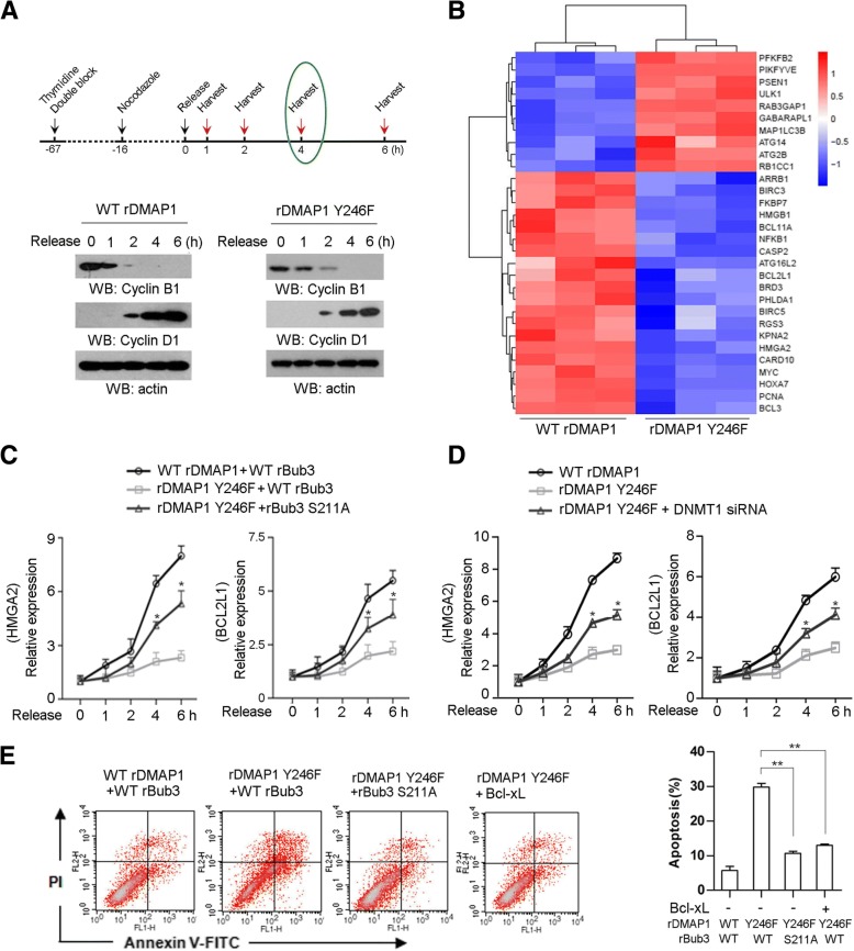
Background: Chromatin modification at mitosis is closely related to transcriptional reactivation in the subsequent cell cycle. We reasoned this process is deregulated by oncogenic signals, which would contribute to mitotic stress resistance in pancreatic cancer. Here, we show DMAP1/Bub3 complex mediates mitotic stress-induced cellular apoptosis, while this effect is counteracted by c-Src in pancreatic cancer cells. Our study aims to uncover an unidentified mechanism underlying the distinct response to mitotic stress between normal cells and pancreatic cancer cells.

Methods: The interaction between Bub3 and DMAP1 upon mitotic stress signaling was determined through molecular and cell biological methods. The inhibitory effect of c-Src on DMAP1/Bub3-mediated DNA methylation and gene transcription profile was investigated. The association between c-Src-mediated DMAP1 phosphorylation and paclitaxel activity in vivo and clinicopathologic characteristics were analyzed.

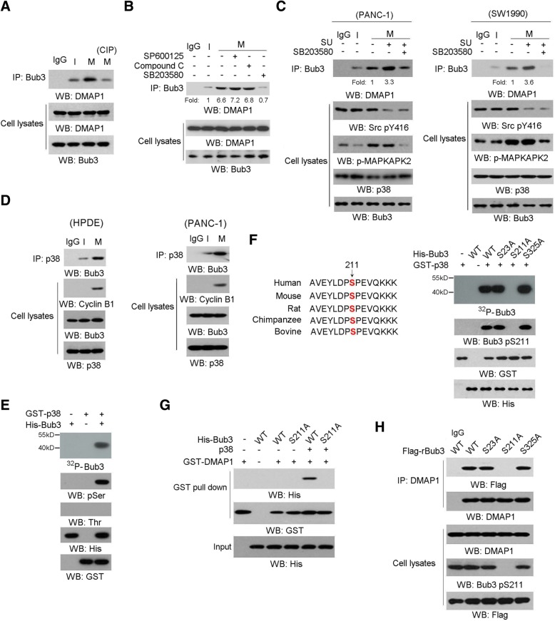
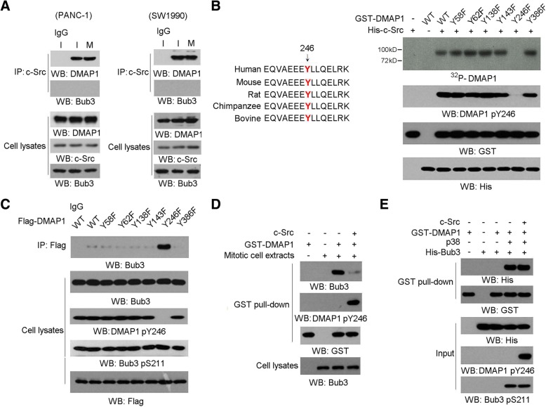
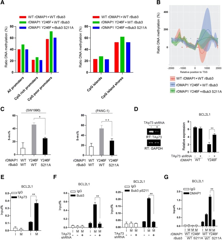
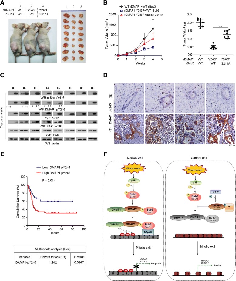
Results: Mitotic arrest induced p38-dependent phosphorylation of Bub3 at Ser211, which promotes DMAP1/Bub3 interaction. DMAP1/Bub3 complex is recruited by TAp73 to the promoter of anti-apoptotic gene BCL2L1, thus mediates the DNA methylation and represses gene transcription linked to cell apoptosis. Meanwhile, DMAP1 was highly phosphorylated at Tyr 246 by c-Src in pancreatic cancer cells, which impedes DMAP1/Bub3 interaction and the relevant cellular activites. Blocking DMAP1 pTyr-246 potentiates paclitaxel-inhibited tumor growth. Clinically, DMAP1 Tyr 246 phosphorylation correlates with c-Src activity in human pancreatic cancer specimens and poor prognosis in pancreatic cancer patients.

Conclusions: Our findings reveal a regulatory role of Bub3 in DMAP1-mediated DNA methylation upon mitotic stress and provide the relevance of DMAP1 pTyr-246 to mitotic stress resistance during pancreatic cancer treatment.

Keywords: Mitosis,transcriptional reactivation,tumourigenesis,Bub3,p38,DMAP1,c-Src.


Conflict of interest statement

Ethics approval and consent to participate

All animal experiments conformed to the guidelines of the Institutional Animal Care and Use Committee of Shanghai Jiaotong University. The use of human tissues was approved by the Institutional Ethics Board of Shanghai General Hospital and conforms to the Helsinki Declaration and to local legislation.

Consent for publication

Not applicable.

Competing interests

The authors declare that they have no competing interests.

Publisher’s Note

Springer Nature remains neutral with regard to jurisdictional claims in published maps and institutional affiliations.

Figures

Fig. 1
Fig. 1
Bub3 interacts with DMAP1 during mitotic arrest. In A-F, immunoblotting analyses were performed using the indicated antibodies; Data represent 1 out of 3 experiments. a, HPDE cells expressing Flag-Bub3 were synchronized in interphase by thymidine double block (2 mM) or were synchronized in mitosis by nocodazole (200 nM) treatment for 16 h after releasing thymidine double block for 8 h (left panel), then cells were extracted and subjected to immunoprecipitation with an anti-Flag antibody. The precipitates from immunoprecipitated-Flag-bub3 were washed by Flag peptides and then were analyzed by coomassie brilliant blue staining and immunoblotting (right panel). b, HPDE cells were synchronized by thymidine double block (2 mM) and were released for 8 h, followed by nocodazole (200 nM) treatment for 16 h, cellular extracts were subjected to immunoprecipitation with an anti-Bub3 antibody. c, HPDE cells were transfected with or without DMAP1 siRNA. Cellular extracts were subjected to immunoprecipitation with an anti-Bub3 antibody. d, HPDE and PANC-1 cells were synchronized by thymidine double block (2 mM) and were released for 8 h, followed by nocodazole (200 nM) treatment for 16 h (left panel). PANC-1 cells were treated with SU6656 (shown as ‘SU’) (10 μM) for 1 h post Nocodazole treatment for 16 h. Cellular extracts were subjected to immunoprecipitation with an anti-Bub3 antibody. e, HPDE cells overexpressed with Src Y527F were synchronized by thymidine double block (2 mM) and were released for 8 h, followed by nocodazole (200 nM) treatment for 16 h (left panel). Cellular extracts subjected to immunoprecipitation with an anti-Bub3 antibody. f, HPDE cells synchronized by thymidine double block (2 mM) were released for 8 h, followed by nocodazole (200 nM) treatment for 16 h (left panel). Cells expressing HA-DMAP1was transfected with plasmids expressing indicated length of Flag-Bub3. Cellular extracts were subjected to immunoprecipitation with an anti-Flag antibody
Fig. 2
Fig. 2
p38 phosphorylates Bub3 at Ser211 and promotes Bub3/DMAP1interaction. In a-h, immunoblotting analyses were performed using the indicated antibodies. Data represent 1 out of 3 experiments. In a-d and h, cells were synchronized in interphase (I) by thymidine (2 mM) double block or were synchronized in mitosis (M) by nocodazole (200 nM) treatment for 16 h after releasing thymidine double block for 8 h. In a-h, data represent 1 out of 3 experiments. a, HPDE cells were synchronized in interphase or mitosis. Cellular extracts were subjected to immunoprecipitation with an anti-Bub3 antibody and the immunoprecipitates were treated with or without CIP (10 units). b, HPDE cells synchronized in interphase or mitosis were treated with Compound C (10 μM), SP600125 (20 μM) and SB203580 (25 μM) for 1 h, after Nocodazole treatment for 16 h. c, PANC-1 or SW1990 cells were synchronized in interphase or mitosis. Cells were treated with SB203580 (25 μM) and SU6656 (shown as ‘SU’) (10 μM) for 1 h, after Nocodazole treatment for 16 h. Cellular extracts were subjected to immunoprecipitation with an anti-Bub3 antibody. d, HPDE or PANC-1 cells were synchronized in interphase or mitosis. Cellular extracts were subjected to immunoprecipitation with an anti-p38 antibody. e and f, In vitro phosphorylation analyses were performed by mixing the purified active p38 with the indicated purified GST-Bub3 proteins in the presence of [γ-32P]ATP. Ser211 of Bub3 is evolutionarily conserved in the indicated species (f, left panel). g, The indicated purified His-Bub3 protein was mixed with GST-DMAP1 purified protein with or without p38. GST pull down analyses were performed. h, HPDE cells synchronized in mitosis were expressed with indicated Flag-Bub3. Cellular extracts were subjected to immunoprecipitation with an anti-Flag antibody
Fig. 3
Fig. 3
c-Src phosphorylates DMAP1 at Tyr246 and disrupts Bub3/DMAP1 complex formation. In a-e, immunoblotting analyses were performed using the indicated antibodies. Data represent 1 out of 3 experiments. In a, c and d, cells were synchronized in interphase (I) by thymidine double block (2 mM) or were synchronized in mitosis (M) by nocodazole (200 nM) treatment for 16 h after releasing thymidine double block for 8 h. In a-e, data represent 1 out of 3 experiments. a, PANC-1 or SW1990 cells were synchronized in interphase or mitosis. Cellular extracts were subjected to immunoprecipitation with an anti-Src antibody. b, In vitro phosphorylation analyses were performed by mixing the purified active c-Src with the indicated purified GST-DMAP1 proteins in the presence of [γ-32P]ATP. Tyr246 of DMAP1 is evolutionarily conserved in the indicated species (b, left panel). c, Cells synchronized in mitosis were expressed with indicated Flag-DMAP1. Cellular extracts were subjected to immunoprecipitation with an anti-Flag antibody Cells. d, The indicated purified GST-DMAP1 protein was mixed with mitotic extracts from PANC-1 cells in the presence or absence of c-Src. GST pull down analyses were performed. e, Purified His-Bub3 protein with or without phosphorylation by c-Src was mixed with GST-DMAP1 purified protein with or without p38. GST pull down analyses were performed
Fig. 4
Fig. 4
Bub3/DMAP1 complex represses anti-apoptotic genes transcription. In a, immunoblotting analyses were performed using the indicated antibodies; data represent 1 out of 3 experiments. In c-e, the values represent mean ± s.e.m. of three independent experiments. a, SW1990 cells were double blocked by thymide and treated with nocodazole (200 nM) following by releasing for the indicated periods. b, SW1990 cells were released for 4 h after thymidine double block and nocodazole (200 nM) for 16 h. Hierachical clustering of 4307 probe sets correlating with DMAP1 Y246F-expressed cells show that genes relevant to anti-apoptosis or autophagy were effective in separating cases from DMAP1 WT-expressed cells. c and d SW1990 cells expressed with the indicated plasmids were treated with nocodazole (200 nM) post thymidine double block, and were released for the indicated time. Relative mRNA levels were analyzed by real-time PCR. In c, * represents p < 0.05 between groups of cells expressing rDMAP1 Y246F plus WT rBub3 and groups of cells expressing rDMAP1 Y246F plus rBub3 S211A. In d, * represents p < 0.05 between groups of cells expressing rDMAP1 Y246F and groups of cells expressing rDMAP1 Y246F plus DNMT1 siRNA. e, Cell apoptosis was analyzed by Annexin V assays followed by flow cytometry. ** represents p < 0.01 between indicated groups
Fig. 5
Fig. 5
c-Src-mediated DMAP1 phosphorylation blocks DMAP1-mediated DNA methylation. a, SW1990 cells expressed with the indicated plasmids were synchronized in mitosis (M) by nocodazole (200 nM) treatment for 16 h after releasing thymidine double block for 8 h. DNA methylation levels of promoters and CpG islands or CpG islands shores were presented as ratio of methylated reads to unmethylated reads. The values represent from 2 repeated samples. b, SW1990 cells expressed with the indicated plasmids were synchronized in mitosis (M) by nocodazole (200 nM) treatment. DNA methylation profile of the promoter region (TSS ±1 kb) of BCL2L1. The values represent from 2 repeated samples. c, SW1990 and PANC-1 cells expressed with the indicated plasmids were synchronized in mitosis (M) by nocodazole (200 nM) treatment. 5-mc levels were examined by bisulfite sequencing using primers covering TAp73 binding site of BCL2L1 gene promoter region. d-g, Cells were synchronized in interphase (I) by thymidine (2 mM) double block or were synchronized in mitosis (M) by nocodazole (200 nM) treatment. d, SW1990 cells with depleted DMAP1, and reconstituted expression of WT rDMAP1 or rDMAP1 Y246F were transfected with plasmid for expression of TAp73 shRNA. Relative mRNA level was analyzed by real-time PCR. e, ChIP analyses were performed in SW1990 cells. The primers covering TAp73 binding site of BCL2L1 gene promoter region were used for the real-time PCR. f, SW1990 cells were transfected with plasmid for expression of TAp73 shRNA. ChIP analyses were performed. The primers covering TAp73 binding site of BCL2L1 gene promoter region were used for the real-time PCR. g, SW1990 cells were expressed with the indicated plasmids. ChIP analyses were performed. The primers covering TAp73 binding site of BCL2L1 gene promoter region were used for the real-time PCR. The y axis shows the value normalized to the input. The values represent mean ± s.e.m. of three independent experiments;*represents p < 0.05 and **represents p < 0.01 between indicated groups
Fig. 6
Fig. 6
DMAP1 Y246 phosphorylation is required for tumourigenesis. a and b, A total of 5 × 106 SW1990 cells with Bub3 or DMAP1 depletion and reconstituted expression of the WT rBub3/WT rDMAP1, WT rBub3/rDMAP1 Y246F or rBub3 S211A/rDMAP1 Y246F were subcutaneously injected into the athymic nude mice. Paclitaxel (5 mg/kg) was injected intraperitoneally every two days once the volume of tumours reached 200 mm3. Data represent the mean ± s.e.m. (n = 8). *represents p < 0.05 and **represents p < 0.01 between indicated groups. Representative tumour xenografts were shown (a). Tumour volumes and weight were measured. In the measurement of tumour volumes, length (a) and width (b) and calculated using the following equation: V = ab2/2. Data represent the means ± s.e.m. (n = 8, right panel) (b). c and d Immunoblotting analyses (c) and Immunohistochemical staining (d) with anti-DMAP1 pY246 was performed on human pancreas tumour specimens. Representative photos were shown. For immunoblotting analyses, the numbers underneath the blotting bands represent the normalized density quantified by using densitometry using Image J 2 × software. e, The survival times for 90 patients with low (0–2 staining scores, blue curve) versus high (2.1–5 staining scores, red curve) DMAP1 Tyr246 phosphorylation (low, 29 patients; high, 61 patients) (upper right) were compared. The Kaplan-Meier method and log-rank tests indicating the significance level of the association of DMAP1 Tyr246 phosphorylation (p = 0.014) with patient survival. The table (lower) shows the cox-multivariate analysis after adjustment for patient sex and age, indicating the significance level of the association of DMAP1 Tyr246 phosphorylation (p = 0.0247, HR = 1.942) with patient survival. f, The schematic diagram showing the regulatory role of Bub3/DMAP1 complex in DNA methylation upon mitotic stress in normal pancreatic epithelial cells and pancreatic cancer cells. Mitotic arrest induces p38 activation, which phosphorylates Bub3 at Ser 211 and promotes its interaction with DMAP1/DNMT1. TAp73 recruits Bub3/DMAP1 complex to promoter regions of its-targeted genes, where DMAP1/DNMT1 mediates DNA methylation and blocks expression of genes responsible for anti-apoptosis. c-Src can phosphorylate DMAP1 at Tyr246, which disrupts Bub3/DMAP1 complex formation. In normal cells with limited c-Src activity, p38/Bub3/DMAP1 signaling is readily activated under mitotic arrest; in turn, cells are prone to apoptosis (left part). In cancer cells, hyperactive c-Src leads to DMAP1 tyr246 phosphorylation and inhibits mitotic arrest-induced cell apoptosis mediated by p38/Bub3/DMAP1 signaling (right part)

References

    1. McIntosh JR. Mitosis. Cold Spring Harb Perspect Biol. 2016;8:a023218. - PMC - PubMed
    1. Musacchio A, Salmon ED. The spindle-assembly checkpoint in space and time. Nat Rev Mol Cell Biol. 2007;8:379–393. doi: 10.1038/nrm2163. - DOI - PubMed
    1. Musacchio A, Hardwick KG. The spindle checkpoint: structural insights into dynamic signalling. Nat Rev Mol Cell Biol. 2002;3:731–741. doi: 10.1038/nrm929. - DOI - PubMed
    1. Vitale I, Galluzzi L, Castedo M, Kroemer G. Mitotic catastrophe: a mechanism for avoiding genomic instability. Nat Rev Mol Cell Biol. 2011;12:385–392. doi: 10.1038/nrm3115. - DOI - PubMed
    1. Strzyz P. Cell signalling: Signalling to cell cycle arrest. Nat Rev Mol Cell Biol. 2016;17:536. doi: 10.1038/nrm.2016.108. - DOI - PubMed

Publication types

MeSH terms