. 2019 Mar:188:369-379.
doi: 10.1016/j.neuroimage.2018.12.027. Epub 2018 Dec 13.

Photoacoustic microscopy of obesity-induced cerebrovascular alterations

Affiliations

Photoacoustic microscopy of obesity-induced cerebrovascular alterations

Rui Cao et al. Neuroimage. 2019 Mar.

Abstract

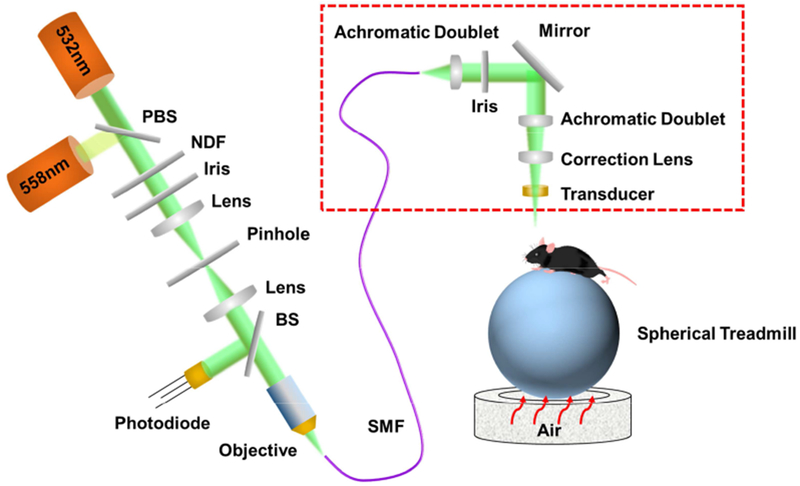
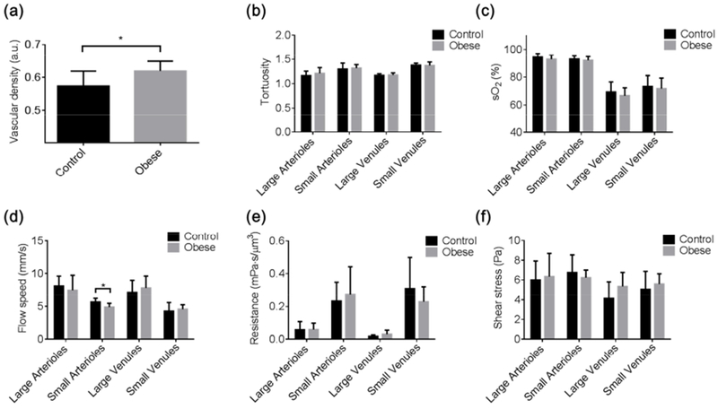
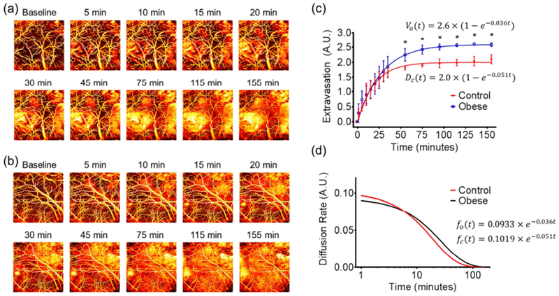
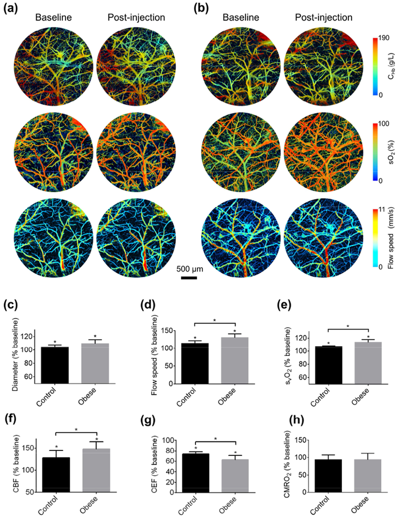
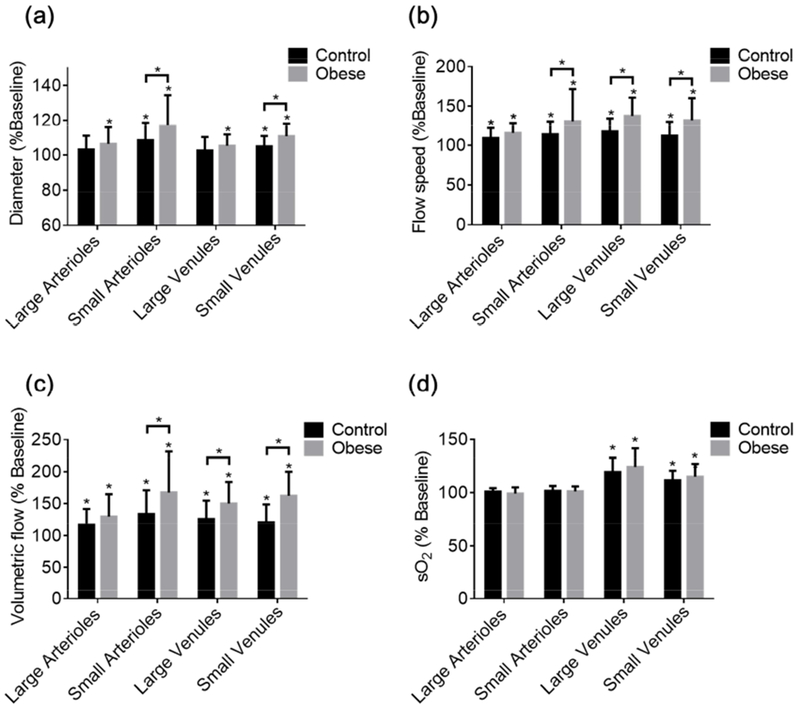
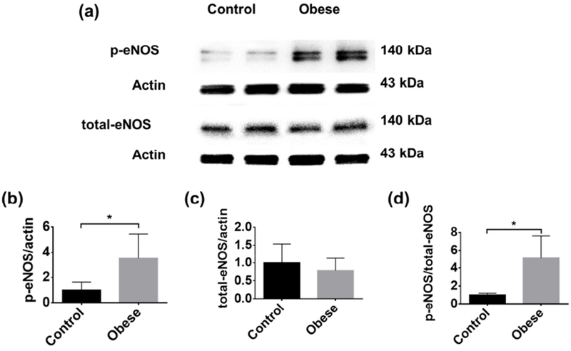
Cerebral small vessel disease has been linked to cognitive, psychiatric and physical disabilities, especially in the elderly. However, the underlying pathophysiology remains incompletely understood, largely due to the limited accessibility of these small vessels in the live brain. Here, we report an intravital imaging and analysis platform for high-resolution, quantitative and comprehensive characterization of pathological alterations in the mouse cerebral microvasculature. By exploiting multi-parametric photoacoustic microscopy (PAM), microvascular structure, blood perfusion, oxygenation and flow were imaged in the awake brain. With the aid of vessel segmentation, these structural and functional parameters were extracted at the single-microvessel level, from which vascular density, tortuosity, wall shear stress, resistance and associated cerebral oxygen extraction fraction and metabolism were also quantified. With the use of vasodilatory stimulus, multifaceted cerebrovascular reactivity (CVR) was characterized in vivo. By extending the classic Evans blue assay to in vivo, permeability of the blood-brain barrier (BBB) was dynamically evaluated. The utility of this enabling technique was examined by studying cerebrovascular alterations in an established mouse model of high-fat diet-induced obesity. Our results revealed increased vascular density, reduced arterial flow, enhanced oxygen extraction, impaired BBB integrity, and increased multifaceted CVR in the obese brain. Interestingly, the 'counterintuitive' increase of CVR was supported by the elevated active endothelial nitric oxide synthase in the obese mouse. Providing comprehensive and quantitative insights into cerebral microvessels and their responses under pathological conditions, this technique opens a new door to mechanistic studies of the cerebral small vessel disease and its implications in neurodegeneration and stroke.

Keywords: Blood-brain barrier; Cerebrovascular reactivity; Obesity; Photoacoustic microscopy; Single-vessel analysis.


Figures

Figure 1.
Figure 1.. Schematic of the head-restrained multi-parametric PAM for imaging the awake mouse brain.
Red dashed box: PAM scan head. PBS, polarizing beam splitter; NDF, neutral density filter; BS, beam sampler; SMF, single-mode fiber.
Figure 2.
Figure 2.. Obesity-induced changes in cerebrovascular structure and function.
PAM-measured, segmentation-enabled quantification of cerebrovascular alterations in the (a) vascular density, (b) vessel tortuosity, (c) sO2, (d) blood flow speed, (e) vascular resistance, and (f) wall shear stress in control (n=6) and obese (n=8) mice. All data are presented as mean ± standard deviation. *, p<0.05.
Figure 3.
Figure 3.. Obesity-induced change in cerebrovascular permeability.
Time-lapse monitoring of Evans blue extravasation in the brains of (a) control and (b) obese mice. (c) Statistical comparison of the dye extravasation dynamics in the control and obese mice (n=5 for each group). (d) Comparison of the dye extravasation rate in the control and obese mice. All data are presented as mean ± standard deviation. *, p<0.05.
Figure 4.
Figure 4.. Obesity-induced change in cerebrovascular reactivity.
Blood perfusion, oxygenation and flow acquired in the brains of (a) control and (b) obese mice before and after tail-vein injection of acetazolamide. Acetazolamide-induced changes in the (c) vessel diameter, (d) blood flow speed, (e) venous sO2, (f) OEF, (g) CBF, and (h) CMRO2 in the control (n=6) and obese (n=8) mice. All data are presented as mean ± standard deviation. *, p<0.05.
Figure 5.
Figure 5.. Vessel type-specific changes in cerebrovascular reactivity.
Acetazolamide-induced changes in the (a) diameter, (b) blood flow speed, (c) volumetric flow, and (d) sO2 of different types of vessels in the control and obese mouse brains. Sample size: 13 large arterioles, 45 small arterioles, 31 large venules, and 22 small venules in the control group and 17 large arterioles, 52 small arterioles, 35 large venules, and 28 small venules in the obese group. All data are presented as mean ± standard deviation, *, p<0.05.
Figure 6.
Figure 6.. Comparison of the eNOS levels in the control and obese mouse brains.
(a) Representative western blot images of phospho-eNOS and total eNOS. Statistical comparison of (b) phospho-eNOS, (c) total eNOS, and (d) the ratio of phospho-eNOS to total eNOS in the control (n=8) and obese (n=8) mouse brains. All data are presented as mean ± standard deviation. *, p<0.05.

References

    1. Acharya NK, Goldwaser EL, Forsberg MM, Godsey GA, Johnson CA, Sarkar A, DeMarshall C, Kosciuk MC, Dash JM, Hale CP, Leonard DM, Appelt DM, Nagele RG, 2015. Sevoflurane and Isoflurane induce structural changes in brain vascular endothelial cells and increase blood-brain barrier permeability: Possible link to postoperative delirium and cognitive decline. Brain Res. 1620, 29–41. doi:10.1016/j.brainres.2015.04.054 - DOI - PubMed
    1. Banerjee G, Wilson D, Jäger HR, Werring DJ, 2016. Novel imaging techniques in cerebral small vessel diseases and vascular cognitive impairment. Biochim. Biophys. Acta - Mol. Basis Dis. 1862, 926–938. doi:10.1016/j.bbadis.2015.12.010 - DOI - PubMed
    1. Blair GW, Doubal FN, Thrippleton MJ, Marshall I, Wardlaw JM, 2016. Magnetic resonance imaging for assessment of cerebrovascular reactivity in cerebral small vessel disease: A systematic review. J. Cereb. Blood Flow Metab. 36, 833–841. doi:10.1177/0271678X16631756 - DOI - PMC - PubMed
    1. Blair GW, Hernandez MV, Thrippleton MJ, Doubal FN, Wardlaw JM, 2017. Advanced Neuroimaging of Cerebral Small Vessel Disease. Curr. Treat. Options Cardiovasc. Med. 19, 56. doi:10.1007/s11936-017-0555-1 - DOI - PMC - PubMed
    1. Blüher M, 2017. Obesity: The myth of innocent obesity. Nat. Rev. Endocrinol doi:10.1038/nrendo.2017.146 - DOI - PubMed

Publication types