Review
. 2019 Jan:105:213-223.
doi: 10.1016/j.molimm.2018.09.026. Epub 2018 Dec 13.

Innate T cells in the intensive care unit

Affiliations
Review

Innate T cells in the intensive care unit

Edy Yong Kim et al. Mol Immunol. 2019 Jan.

Abstract

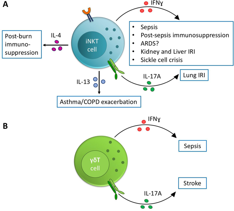
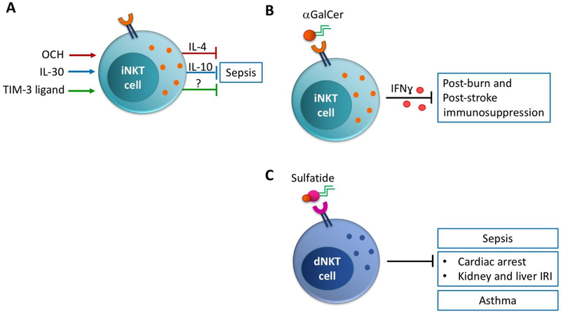
Rapid onset of acute inflammation is a hallmark of critical illnesses that bring patients to the intensive care unit (ICU). In critical illness, innate T cells rapidly reach full activation and drive a robust acute inflammatory response. As "cellular adjuvants," innate T cells worsen inflammation and mortality in several common critical illnesses including sepsis, ischemia-reperfusion injury, stroke, and exacerbations of respiratory disease. Interestingly, innate T cell subsets can also promote a protective and anti-inflammatory response in sepsis, ischemia-reperfusion injury, and asthma. Therapies that target innate T cells have been validated in several models of critical illness. Here, we review the role of natural killer T (NKT) cells, mucosal-associated invariant T (MAIT) cells and γδ T cells in clinical and experimental critical illness.

Keywords: Cardiac arrest; Critical care; Gamma delta T cell; MAIT cell; NKT cell; Sepsis.


Figures

Figure 1.
Figure 1.. Innate T cells drive critical illness.
(A) Invariant NKT (iNKT) cells drive the production of inflammatory and immunosuppressive cytokines that worsen outcomes in experimental critical illnesses. IRI = ischemia-reperfusion injury. ARDS = acute respiratory distress syndrome. (B) γδ T cells drive the production of inflammatory cytokines that worsen outcomes in experimental sepsis and stroke.
Figure 2.
Figure 2.. Innate T cells are therapeutic targets in critical illness.
(A) Treatment with altered lipid antigens (OCH), exogenous cytokines (IL-30) or ligands for immune checkpoint receptors (Tim-3) skew iNKT cells towards an anti-inflammatory phenotype and improve outcomes in experimental sepsis. (B) αGalCer antigen activates iNKT cells, which coordinate production of IFNγ and ameliorate post-burn and post-stroke immunosuppression. (C) Sulfatide antigen activates diverse NKT cells, which improve outcomes in experimental sepsis, ischemia-reperfusion injury (IRI) and asthma exacerbation.

Similar articles

Cited by

References

    1. Akbari O, Stock P, Meyer E, Kronenberg M, Sidobre S, Nakayama T, Taniguchi M, Grusby MJ, DeKruyff RH, Umetsu DT, 2003. Essential role of NKT cells producing IL-4 and IL-13 in the development of allergen-induced airway hyperreactivity. Nat. Med 9, 582–588. 10.1038/nm851 - DOI - PubMed
    1. Aoyagi T, Yamamoto N, Hatta M, Tanno D, Miyazato A, Ishii K, Suzuki K, Nakayama T, Taniguchi M, Kunishima H, Hirakata Y, Kaku M, Kawakami K, 2011. Activation of pulmonary invariant NKT cells leads to exacerbation of acute lung injury caused by LPS through local production of IFN-gamma and TNF-alpha by Gr-1+ monocytes. Int Immunol 23, 97–108. 10.1093/intimm/dxq460 - DOI - PubMed
    1. Arrenberg P, Maricic I, Kumar V, 2011. Sulfatide-mediated activation of type II natural killer T cells prevents hepatic ischemic reperfusion injury in mice. Gastroenterology 140, 646–655. 10.1053/j.gastro.2010.10.003 - DOI - PMC - PubMed
    1. Bai L, Picard D, Anderson B, Chaudhary V, Luoma A, Jabri B, Adams EJ, Savage PB, Bendelac A, 2012. The majority of CD1d-sulfatide-specific T cells in human blood use a semiinvariant Vdelta1 TCR. Eur J Immunol 42, 2505–2510. 10.1002/eji.201242531 - DOI - PMC - PubMed
    1. Beaudoin L, Laloux VE, Novak J, Lucas B, Lehuen A, 2002. NKT cells inhibit the onset of diabetes by impairing the development of pathogenic T cells specific for pancreatic β cells. Immunity 17, 725–736. 10.1016/S1074-7613(02)00473-9 - DOI - PubMed

Publication types

Substances