. 2019 Feb 15;294(7):2470-2485.
doi: 10.1074/jbc.RA118.004836. Epub 2018 Dec 15.

A serine in the first transmembrane domain of the human E3 ubiquitin ligase MARCH9 is critical for down-regulation of its protein substrates

Affiliations

A serine in the first transmembrane domain of the human E3 ubiquitin ligase MARCH9 is critical for down-regulation of its protein substrates

Cyrus Tan et al. J Biol Chem. .

Abstract

The membrane-associated RING-CH (MARCH) family of membrane-bound E3 ubiquitin ligases regulates the levels of cell-surface membrane proteins, many of which are involved in immune responses. Although their role in ubiquitin-dependent endocytosis and degradation of cell-surface proteins is extensively documented, the features of MARCH proteins and their substrates that drive the molecular recognition events leading to ubiquitin transfer remain poorly defined. In this study, we sought to determine the features of human MARCH9 that are required for regulating the surface levels of its substrate proteins. Consistent with previous studies of other MARCH proteins, we found that susceptibility to MARCH9 activity is encoded in the transmembrane (TM) domains of its substrates. Accordingly, substitutions at specific residues and motifs within MARCH9's TM domains resulted in varying degrees of functional impairment. Most notably, a single serine-to-alanine substitution in the first of its two TM domains rendered MARCH9 completely unable to alter the surface levels of two different substrates: the major histocompatibility class I molecule HLA-A2 and the T-cell co-receptor CD4. Solution NMR analysis of a MARCH9 fragment encompassing the two TM domains and extracellular connecting loop revealed that the residues contributing most to MARCH9 activity are located in the α-helical portions of TM1 and TM2 that are closest to the extracellular face of the lipid bilayer. This observation defines a key region required for substrate regulation. In summary, our biochemical and structural findings demonstrate that specific sequences in the α-helical MARCH9 TM domains make crucial contributions to its ability to down-regulate its protein substrates.

Keywords: E3 ligase; T-cell; immunology; major histocompatibility complex (MHC); membrane protein; membrane-associated RING-CH (MARCH); nuclear magnetic resonance (NMR); receptor endocytosis; transmembrane domain; ubiquitin ligase.


Conflict of interest statement

The authors declare that they have no conflicts of interest with the contents of this article

Figures
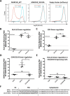
Figure 1.
Figure 1.
Susceptibility to MARCH9-mediated down-regulation can be transferred by the CD4 TM domain sequence. A, representative histograms showing 293T cells transduced with lentivirus-encoding dox-inducible WT (left), W143A RING mutant (middle), or no MARCH9 (right) with a constitutively expressed mCherry marker. Transduced cells were incubated with or without dox for 48 h, and surface levels of endogenous HLA-A2 on mCherry+ cells were measured by flow cytometry. B and C, 293T cells stably expressing human CD4 were transduced with lentiviral constructs shown in A, and surface levels of HLA-A2 in B and CD4 in C were measured on mCherry+ cells after 48 h of culture with or without dox. Data are presented as percentage of substrate remaining on the surface of dox-treated cells relative to untreated cells from the same transduction. Dashed lines indicate mean substrate level remaining on dox-treated cells expressing control WT (lower) and RING mutant W143A (upper) MARCH9. D and E, 293T cells stably expressing human CD4, human CD19, or a chimeric CD19 protein containing the TM domain of CD4 (CD19–CD4TM) were transduced with WT MARCH9 and cultured with and without doxycycline for 48 h. Surface levels of CD4/CD19 (D) and HLA-A2 (E) were measured on mCherry+ cells by flow cytometry and presented as in B and C. In all graphs, lines represent the mean and S.E. of three independent experiments performed on different days (n = 3), and each point represents data from one experiment. Unpaired t test: ns, not significant; **, p < 0.02. F, human CD4 and CD19 partial amino acid sequences showing the residues that were swapped in the CD19–CD4TM chimera. Predicted TM domains underlined. Lysine residues in cytoplasmic tails highlighted in bold represent possible ubiquitination sites.
Figure 2.
Figure 2.
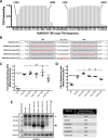
MARCH9 TM sequences contain features that are absolutely required for substrate down-regulation. A, TMHMM (46) prediction of the locations of two TM domains in human MARCH9. Shaded bars are 50% confidence or better, and boundary residues for TM1 (Val-183–Ser-205) and TM2 (Leu-218–His-240) are marked above the graph. B, sequences of mutants analyzed in this figure. Red bold font indicates substituted amino acids. Two cysteines in predicted TM2 were not substituted. MARCH9_ScramLoop was generated by randomizing the 12-amino acid loop sequence such that no position contained the native amino acid but the overall amino acid content was unchanged. C and D, 293T cells stably expressing human CD4 were transduced with the indicated lentiviral constructs, and surface levels of HLA-A2 (C) and CD4 (D) were measured after 48 h culture with or without dox as in Fig. 1. Dashed lines indicate mean substrate level remaining on dox-treated cells expressing control WT (lower) and RING mutant W143A (upper) MARCH9. Unpaired t test: ns, not significant; *, p < 0.05. E, Western blot analysis of MARCH9 expression. WT and mutant MARCH9 sequences with C-terminal HA tag were transiently expressed in 293T cells under the strong EF1α promoter by calcium phosphate transfection and harvested 24–48 h later. Whole-cell lysates were separated by reducing SDS-PAGE, transferred to PVDF, and sequentially immunoblotted using α-GAPDH and α-HA antibodies. + marks the positions of a cellular product detected by SA-HRP; arrow marks the position of the HA-tagged MARCH9 protein. Table shows expression normalized to GAPDH loading control and relative to MARCH9-WT, quantitated by densitometry.
Figure 3.
Figure 3.
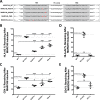
Serine 198 in the first predicted TM domain of MARCH9 is absolutely required for substrate down-regulation. A, sequences of mutants analyzed in this figure. Red bold font indicates substituted amino acids. Vertical box highlights the position of Ser-198. B–E, 293T cells stably expressing human CD4 were transduced with the indicated lentiviral constructs, and surface levels of HLA-A2 (B and D) and CD4 (C and E) were measured after 48 h of culture with or without dox as in Fig. 1. Serine-to-alanine substitutions were examined in blocks of three or four (3S-A and 4S-A mutants; B and C) and separately for the single-serine mutants as indicated (D and E). Dashed lines indicate mean substrate level remaining on dox-treated cells expressing control WT (lower) and RING mutant W143A (upper) MARCH9. Unpaired t test: ns, not significant. F, Western blot analysis of MARCH9 WT and inactive mutant expression, performed as in Fig. 2E. + marks the position of a cellular product detected by SA-HRP; arrow marks the position of MARCH9 protein. Table shows expression normalized to GAPDH loading control and relative to MARCH9-WT, quantitated by densitometry.
Figure 4.
Figure 4.
Serine 198 mutations do not alter MARCH9 subcellular localization. Representative images are shown for 293T cells expressing free cytosolic ZsGreen protein (empty vector, for reference) or MARCH9–ZsGreen fusion proteins with the indicated mutations. Cell preparations were stained and imaged by confocal microscopy 48 h after transient transfection. Color channels are ZsGreen (yellow), F-actin stain (Alexa 555-phalloidin, magenta), and nuclear stain (DAPI, cyan). All images contain both transfected and untransfected cells.
Figure 5.
Figure 5.
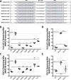
Mutation of tryptophans in TM1 of MARCH9 causes moderate defects, but mutation of a glycophorin A-like motif does not. A, sequences of mutants analyzed in this figure. Red bold font indicates substituted amino acids. B–E, 293T cells stably expressing human CD4 were transduced with the indicated lentiviral constructs, and surface levels of HLA-A2 (B and D) and CD4 (C and E) were measured after 48 h culture with or without dox as in Fig. 1. Dashed lines indicate mean substrate level remaining on dox-treated cells expressing control WT (lower) and RING mutant W143A (upper) MARCH9. Unpaired t test: ns, not significant; *, p < 0.05; **, p < 0.02; ***, p < 0.001; ****, p < 0.0001.
Figure 6.
Figure 6.
Mutation of small, polar, and aromatic residues in TM2 of MARCH9 affects substrate down-regulation. A, sequences of mutants analyzed in this figure. Red bold font indicates substituted amino acids. B–E, 293T cells stably expressing human CD4 were transduced with the indicated lentiviral constructs, and surface levels of HLA-A2 (B and D) and CD4 (C and E) were measured after 48 h of culture with or without dox as in Fig. 1. Dashed lines indicate mean substrate level remaining on dox-treated cells expressing control WT (lower) and inactive mutant S198A (upper) MARCH9. Unpaired t test: ns, not significant; *, p < 0.05; **, p < 0.02; ***, p < 0.001; ****, p < 0.0001.
Figure 7.
Figure 7.
Relationship between MARCH9 mutant activity and protein levels. Expression levels for the indicated mutants (y axis) were analyzed by transient transfection in 293T cells as shown in Figs. 2 and 3, and MARCH9-specific bands were quantitated by densitometry. MARCH9 expression was normalized to GAPDH signal and plotted relative to WT (open circle). Activity score (x axis) was determined by the reduction in surface levels of HLA-A2 after dox treatment (data shown in Figs. 2–5) and expressed as a percentage of WT activity. Dashed line represents expression of WT MARCH9, which was set to 1 for comparison. Vertical error bars represent S.D. of at least three independent transfections (n ≥ 3). Horizontal error bars represent S.E. of three independent experiments (n = 3). Red symbols highlight mutants for which functional defects are likely to be independent of total cellular protein levels.
Figure 8.
Figure 8.
Structural analysis of a MARCH9 TM–loop–TM fragment by solution NMR. A, assigned 1H–15N HSQC spectrum of a uniformly 15N,13C,2H 80%-labeled, 65-amino acid MARCH9 TM–loop–TM peptide (1 mm) in 250 mm LMPG, 20 mm phosphate buffer, pH 6.8, 5% D2O at 600 MHz and 40 °C. The TM–loop–TM peptide contained mutations C223S, M226V, M230V, and C234S to aid in production and an I239V substitution that is present in the mouse sequence (see “Experimental procedures”). B, secondary structure prediction from assigned backbone chemical shifts using TALOS+ (49). Confidence scores for residues predicted to be part of a helix (red bars) or random coil (blue bars) are superimposed with TMHMM predictions (black lines/dots) for comparison. C, backbone torsion angle ϕ (dots) and ψ (triangles) predictions from TALOS+. Values are in degrees, and error bars represent the estimated S.D. of the prediction error. D, backbone 15N T1 (top) and T2 (bottom) relaxation times (milliseconds) determined for a uniformly 15N-labeled MARCH9–TM sample prepared as in A and analyzed at 600 MHz and 40 °C. Helix 1 and Helix 2 are highlighted in large shaded boxes. The positions of key residues Ser-198, Gly-225, and Tyr-227 are highlighted in vertical shaded boxes.

Similar articles

Cited by

References

    1. Bartee E., Mansouri M., Hovey Nerenberg B. T., Gouveia K., and Früh K. (2004) Downregulation of major histocompatibility complex class I by human ubiquitin ligases related to viral immune evasion proteins. J. Virol. 78, 1109–1120 10.1128/JVI.78.3.1109-1120.2004 - DOI - PMC - PubMed
    1. Goto E., Ishido S., Sato Y., Ohgimoto S., Ohgimoto K., Nagano-Fujii M., and Hotta H. (2003) c-MIR, a human E3 ubiquitin ligase, is a functional homolog of herpesvirus proteins MIR1 and MIR2 and has similar activity. J. Biol. Chem. 278, 14657–14668 10.1074/jbc.M211285200 - DOI - PubMed
    1. Ohmura-Hoshino M., Goto E., Matsuki Y., Aoki M., Mito M., Uematsu M., Hotta H, and Ishido S. (2006) A novel family of membrane-bound E3 ubiquitin ligases. J. Biochem. 140, 147–154 10.1093/jb/mvj160 - DOI - PubMed
    1. Baravalle G., Park H., McSweeney M., Ohmura-Hoshino M., Matsuki Y., Ishido S., and Shin J.-S. (2011) Ubiquitination of CD86 is a key mechanism in regulating antigen presentation by dendritic cells. J. Immunol. 187, 2966–2973 10.4049/jimmunol.1101643 - DOI - PMC - PubMed
    1. Furuta K., Walseng E., and Roche P. A. (2013) Internalizing MHC class II–peptide complexes are ubiquitinated in early endosomes and targeted for lysosomal degradation. Proc. Natl. Acad. Sci. U.S.A. 110, 20188–20193 10.1073/pnas.1312994110 - DOI - PMC - PubMed

Publication types

MeSH terms