. 2019 Feb;211(2):563-578.
doi: 10.1534/genetics.118.301711. Epub 2018 Dec 15.

Nucleosome Positioning by an Evolutionarily Conserved Chromatin Remodeler Prevents Aberrant DNA Methylation in Neurospora

Affiliations

Nucleosome Positioning by an Evolutionarily Conserved Chromatin Remodeler Prevents Aberrant DNA Methylation in Neurospora

Andrew D Klocko et al. Genetics. 2019 Feb.

Abstract

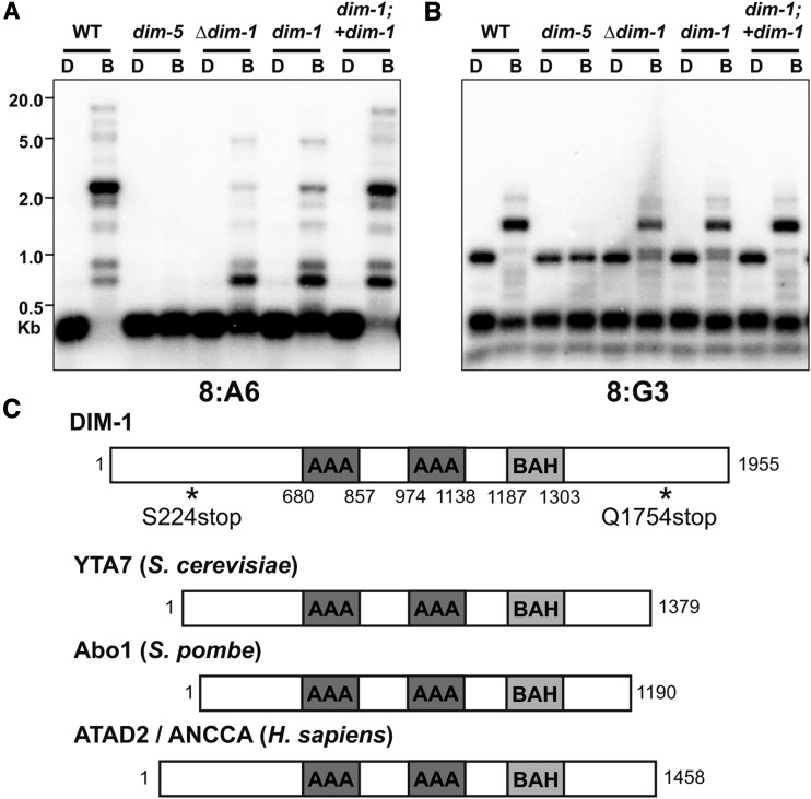
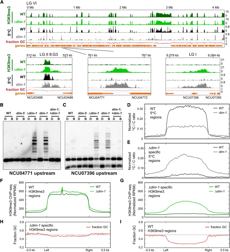
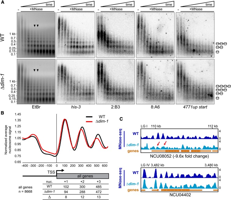
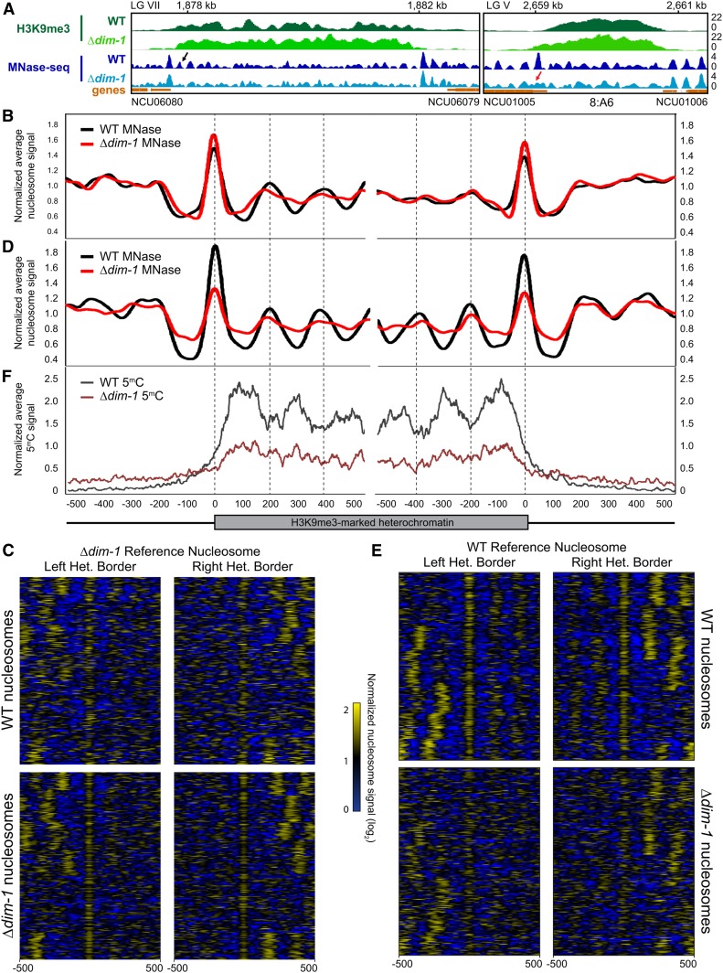
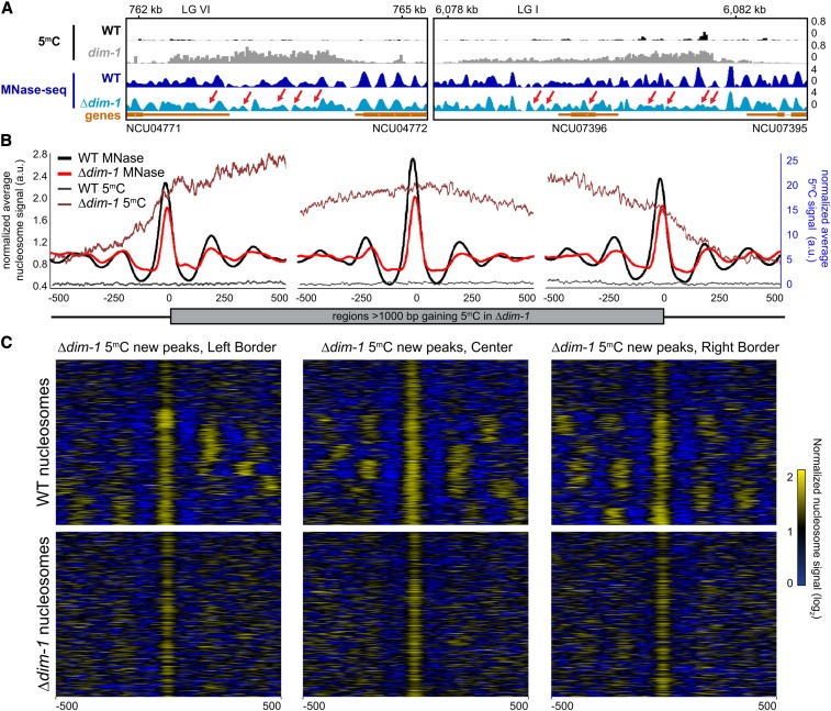
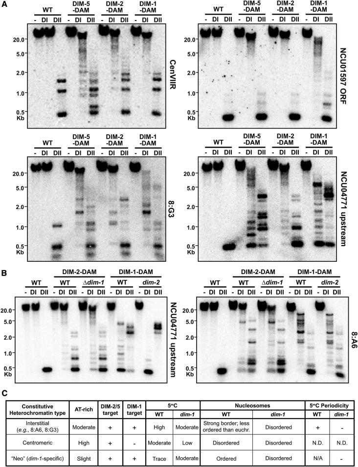
In the filamentous fungus Neurospora crassa, constitutive heterochromatin is marked by tri-methylation of histone H3 lysine 9 (H3K9me3) and DNA methylation. We identified mutations in the Neurospora defective in methylation-1 (dim-1) gene that cause defects in cytosine methylation and implicate a putative AAA-ATPase chromatin remodeler. Although it was well-established that chromatin remodelers can affect transcription by influencing DNA accessibility with nucleosomes, little was known about the role of remodelers on chromatin that is normally not transcribed, including regions of constitutive heterochromatin. We found that dim-1 mutants display both reduced DNA methylation in heterochromatic regions as well as increased DNA methylation and H3K9me3 in some intergenic regions associated with highly expressed genes. Deletion of dim-1 leads to atypically spaced nucleosomes throughout the genome and numerous changes in gene expression. DIM-1 localizes to both heterochromatin and intergenic regions that become hyper-methylated in dim-1 strains. Our findings indicate that DIM-1 normally positions nucleosomes in both heterochromatin and euchromatin and that the standard arrangement and density of nucleosomes is required for the proper function of heterochromatin machinery.

Keywords: CATP; DIM-1; DNA methylation; Neurospora crassa; heterochromatin; nucleosome.


Figures

Figure 1
Figure 1
DNA methylation is abnormal in dim-1 strains at select loci, and DIM-1 has features of ATP-dependent chromatin remodeling factors. (A and B) Southern blot of genomic DNA from the indicated strains digested with either DpnII (D) or its 5mC-sensitive isoschizomer BfuCI (B), and probed for the heterochromatic regions 8:A6 (A) or 8:G3 (B). To test for complementation of the dim-1 mutation, we crossed in a WT allele inserted at the his-3 locus (“+dim-1”) (C). Primary structure schematics of DIM-1 and its homologs from yeasts and humans with known domains are indicated, amino acid coordinates are shown below, and positions of nonsense mutations in dim-1 strains are shown by asterisks.
Figure 2
Figure 2
DNA methylation and H3K9me3 are abnormal in dim-1 strains genome-wide. (A) ChIP-sequencing tracks (merged replicates) for H3K9me3 (green) or bisulfite-sequencing for cytosine methylation (black/gray) for WT or dim-1 strains, and base composition (“fraction GC,” red); all tracks are displayed in 25 bp windows. LG VI is shown, but comparable results were obtained for the other six chromosomes (Figure S4). Selected regions are shown expanded below; different chromosomes [Linkage Groups (LGs)] are identified. Tracks here and in other figures are displayed with the Integrative Genomics Viewer (Robinson et al. 2011) with coordinates relative to the left telomere, and NCU numbers of genes, shown above and below, respectively. (B and C) Southern blots of genomic DNA from the indicated strains digested with either DpnII (D) or its 5mC-sensitive isoschizomer BfuCI (B) and probed for the labeled regions, as in Figure 1A; “upstream” denotes the intergenic region upstream of that gene. (D and E) Average enrichment profiles of bisulfite-sequencing data showing the ratio of methylated-to-unmethylated cytosines across H3K9me3-marked regions in WT strains (D; n = 210) or specifically found in Δdim-1 strains (E; n = 239). Plots show 500 bp upstream and downstream of the left and right borders of constitutive heterochromatic regions, which were scaled to 1 kb for presentation. y-axis denotes the normalized ratio of methylated cytosines/total cytosines per amount of sequencing genome coverage. (F and G) Average enrichment profiles, as in D and E, but of H3K9me3 ChIP-sequencing enrichment in WT and Δdim-1 strains merged from two experiments; y-axis denotes the normalized amount of ChIP-sequencing reads per kilobase per million total reads (RPKM). (H and I) Average enrichment profiles, as in B and C, but of fraction of GC base pairs; horizontal line marks the position where base composition is 50% G+C.
Figure 3
Figure 3
Nucleosome positioning is altered at the TSSs of genes in Δdim-1 strain. (A) Southern blots of DNA from 20-min time course micrococcal nuclease (MNase) digest with WT and Δdim-1 nuclei, probed for the indicated regions; the “NCU04771up start” probe covers the intergenic promoter region of gene NCU04771, including the nucleosome-free region. Arrowheads indicate the time points (8 and 10 min) from which mono- and di-nucleosomes and intervening DNA was purified for paired-end high-throughput sequencing (below). Cartoons at right show interpretation of nucleosome patterns leading to smallest fragments. (B) Average nucleosome enrichment profiles of MNase-sequencing data from WT and Δdim-1 strains normalized to the average signal across the 5′ end of all Neurospora genes, spanning 400 bp upstream to 600 bp downstream of the TSS; numbers below indicate the peak apices, in base pairs from the transcriptional start site (TSS) in WT and Δdim-1 strains, and the difference in the apex position, for the +1, +2, and +3 nucleosomes. (C) Representative examples of nucleosome positions in individual genes with changed (NCU08052), or unchanged (NCU04402; dim-5) expression in a Δdim-1 background. Red arrows highlight nucleosome disorder in a Δdim-1 strain of two nucleosomes that are well-positioned in a WT strain.
Figure 4
Figure 4
Constitutive heterochromatic regions exhibit nucleosome disorder, especially in Δdim-1 strains. (A) Representative examples of nucleosome positioning within constitutive heterochromatin. H3K9me3 ChIP-sequencing (green) and MNase-sequencing (blue) tracks of WT and Δdim-1 strains are shown for a region on LG VII between NCU06080 and NCU06079 and the 8:A6 region on LG V (Selker et al. 2003). The black arrow denotes an internal nucleosome that has periodicity in the WT strain while the red arrow denotes a border nucleosome that becomes disordered in the ?dim-1 strain. (B) Average mono-nucleosome enrichment profiles of MNase-sequencing data (MNase) in WT and Δdim-1 strains at the left and right borders (relative to the left telomere) of constitutive heterochromatic regions longer than at least 1000 base pairs aligned from the first nucleosome peak completely covered by H3K9me3 in Δdim-1 strains. (C) Nucleosome peak signal heatmaps from WT and Δdim-1 strains normalized to the average enrichment signal. Data are shown for ±500 bp for the left and right heterochromatic region borders (n = 210) relative to the first heterochromatic nucleosome peak in Δdim-1 strains. Heterochromatic regions are identically ordered in each heatmap group. (D) Average mono-nucleosome enrichment profiles of MNase-sequencing data (MNase) in WT and Δdim-1 strains at the left and right borders, as in B. Vertical dashed lines mark the apices of WT nucleosome signals. (E) Nucleosome peak signal heatmaps from WT and Δdim-1 strains normalized to the average enrichment signal, as in C, relative to the first heterochromatic nucleosome peak in WT strains. (F) Average enrichment profiles of bisulfite-sequencing showing enrichment of cytosine methylation in WT and Δdim-1 strains at the left and right borders of constitutive heterochromatic regions.
Figure 5
Figure 5
Intergenic regions that gain cytosine methylation in Δdim-1 exhibit aberrant nucleosome positioning. (A) Representative examples of nucleosome positions within intergenic regions that gain cytosine methylation. Tracks of bisulfite-sequencing for WT (black) and dim-1 (light gray), as well as MNase-sequencing for WT (dark blue) and Δdim-1 (light blue) strains are shown. Red arrows highlight changes in nucleosome positioning in Δdim-1 strain. (B) Average mono-nucleosome enrichment profiles (MNase) and cytosine methylation (5mC) in WT and Δdim-1 strains at the first nucleosome apex in left and right borders of regions that gain methylation in a dim-1 strain, as well as a central reference nucleosome peak in a WT strain (n = 322); the WT data used reference nucleosomes in a WT strain while the dim-1 data used reference nucleosomes from a Δdim-1 strain. (C) Heatmaps of nucleosome signals from WT and Δdim-1 strains normalized to average nucleosome signal examining ±500 bp of the intergenic regions gaining cytosine methylation in Δdim-1 strain shown in B; regions are shown in each heatmap pair in the same order.
Figure 6
Figure 6
DIM-1 and heterochromatin machinery localize to heterochromatic regions and intergenic regions. (A) DamID Southern blots of genomic DNA of the indicated dim+ strains digested with DpnI (cuts GAmTC; DI) or its isoschizomer DpnII (inhibited by adenine methylation; DII) or left undigested (−) and probed for the indicated regions. (B) DamID Southern blots, as in A, for different mutant backgrounds expressing the indicated DAM fusion constructs. (C) Table summarizing the phenotypes of a dim-1 strain. N.D., not determined.

Similar articles

Cited by

References

    1. Adhvaryu K. K., Berge E., Tamaru H., Freitag M., Selker E. U., 2011. Substitutions in the amino-terminal tail of Neurospora histone H3 have varied effects on DNA methylation. PLoS Genet. 7: e1002423 10.1371/journal.pgen.1002423 - DOI - PMC - PubMed
    1. Afgan E., Baker D., Batut B., van den Beek M., Bouvier D., et al. , 2018. The Galaxy platform for accessible, reproducible and collaborative biomedical analyses: 2018 update. Nucleic Acids Res. 46: W537–W544. 10.1093/nar/gky379 - DOI - PMC - PubMed
    1. Allshire R. C., Madhani H. D., 2017. Ten principles of heterochromatin formation and function. Nat. Rev. Mol. Cell Biol. 19: 229–244. 10.1038/nrm.2017.119 - DOI - PMC - PubMed
    1. Boussouar F., Jamshidikia M., Morozumi Y., Rousseaux S., Khochbin S., 2013. Malignant genome reprogramming by ATAD2. Biochim. Biophys. Acta 1829: 1010–1014. 10.1016/j.bbagrm.2013.06.003 - DOI - PubMed
    1. Bühler M., Gasser S. M., 2009. Silent chromatin at the middle and ends: lessons from yeasts. EMBO J. 28: 2149–2161. 10.1038/emboj.2009.185 - DOI - PMC - PubMed

Publication types