

Intraoperative near-infrared imaging with receptor-specific versus passive delivery of fluorescent agents in pituitary adenomas
- PMID: 30554181
- PMCID: PMC10985533
- DOI: 10.3171/2018.7.JNS181642
Intraoperative near-infrared imaging with receptor-specific versus passive delivery of fluorescent agents in pituitary adenomas
Abstract
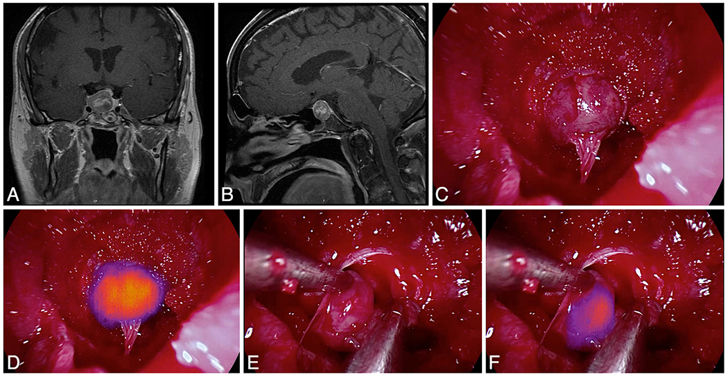
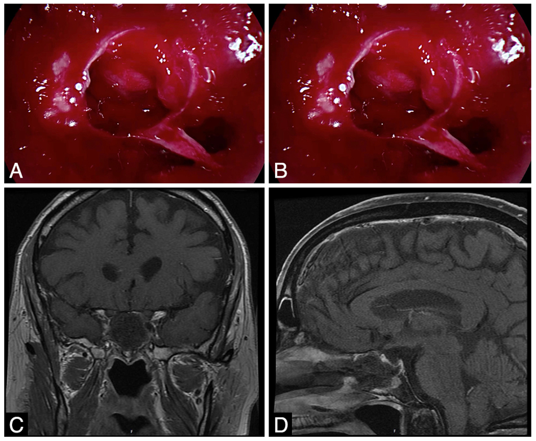
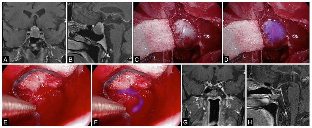
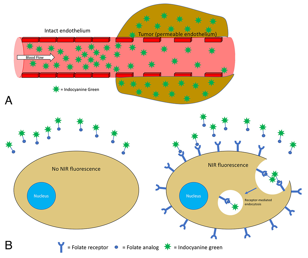
Objective: Intraoperative molecular imaging with tumor-targeted fluorescent dyes can enhance resection rates. In contrast to visible-light fluorophores (e.g., 5-aminolevulinic-acid), near-infrared (NIR) fluorophores have increased photon tissue penetration and less contamination from tissue autofluorescence. The second-window ICG (SWIG) technique relies on passive accumulation of indocyanine green (ICG) in neoplastic tissues. OTL38, conversely, targets folate receptor overexpression in nonfunctioning pituitary adenomas. In this study, we compare the properties of these 2 modalities for NIR imaging of pituitary adenomas to better understand the potential for NIR imaging in neurosurgery.
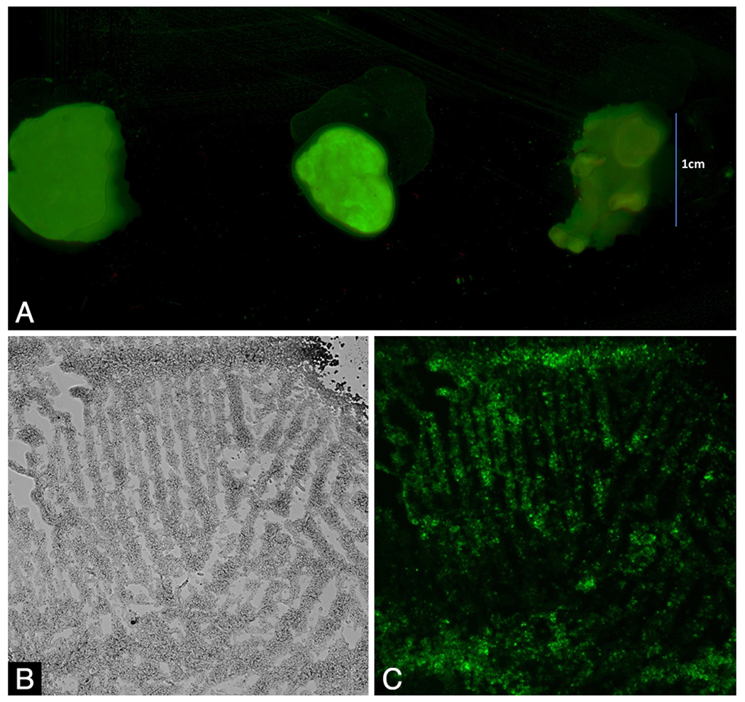
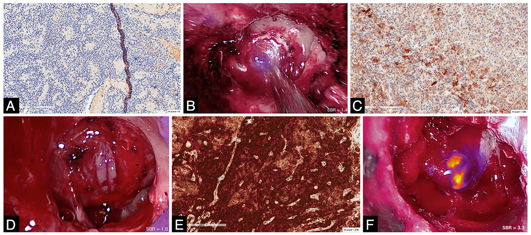
Methods: A total of 39 patients with pituitary adenomas were enrolled between June 2015 and January 2018 in 2, sequential, IRB-approved studies. Sixteen patients received systemic ICG infusions 24 hours prior to surgery, and another 23 patients received OTL38 infusions 2-3 hours prior to surgery. NIR fluorescence signal-to-background ratio (SBR) was recorded during and after resection. Immunohistochemistry was performed on the 23 adenomas resected from patients who received OTL38 to assess expression of folate receptor-alpha (FRα).
Results: All 16 adenomas operated on after ICG administration demonstrated strong NIR fluorescence (mean SBR 4.1 ± 0.69 [SD]). There was no statistically significant difference between the 9 functioning and 7 nonfunctioning adenomas (p = 0.9). After administration of OTL38, the mean SBR was 1.7 ± 0.47 for functioning adenomas, 2.6 ± 0.91 for all nonfunctioning adenomas, and 3.2 ± 0.53 for the subset of FRα-overexpressing adenomas. Tissue identification with white light alone for all adenomas demonstrated 88% sensitivity and 90% specificity. SWIG demonstrated 100% sensitivity but only 29% specificity for both functioning and nonfunctioning adenomas. OTL38 was 75% sensitive and 100% specific for all nonfunctioning adenomas, but when assessment was limited to the 9 FRα-overexpressing adenomas, the sensitivity and specificity of OTL38 were both 100%.
Conclusions: Intraoperative imaging with NIR fluorophores demonstrates highly sensitive detection of pituitary adenomas. OTL38, a folate-receptor-targeted fluorophore, is highly specific for nonfunctioning adenomas but has no utility in functioning adenomas. SWIG, which relies on passive diffusion into neoplastic tissue, is applicable to both functioning and nonfunctioning pituitary adenomas, but it is less specific than targeted fluorophores. Thus, targeted and nontargeted NIR fluorophores play important, yet distinct, roles in intraoperative imaging. Selectively and intelligently using either agent has the potential to greatly improve resection rates and outcomes for patients with intracranial tumors.
Keywords: 5-ALA = 5-aminolevulinic acid; ACTH = adrenocorticotropic hormone; EPR = enhanced permeability and retention; FRα = folate receptor–alpha; GH = growth hormone; ICG = indocyanine green; NIR = near-infrared; NPV = negative predictive value; PPV = positive predictive value; SBR = signal-to-background ratio; SPECT = single-photon emission computed tomography; SWIG = second-window ICG; TSH = thyroid-stimulating hormone; folate receptor; indocyanine-green; near-infrared imaging; pituitary adenoma; pituitary surgery; targeted imaging.
Figures
Similar articles
-
Folate receptor overexpression can be visualized in real time during pituitary adenoma endoscopic transsphenoidal surgery with near-infrared imaging.J Neurosurg. 2018 Aug;129(2):390-403. doi: 10.3171/2017.2.JNS163191. Epub 2017 Aug 25. J Neurosurg. 2018. PMID: 28841122 Free PMC article.
-
Folate Receptor Near-Infrared Optical Imaging Provides Sensitive and Specific Intraoperative Visualization of Nonfunctional Pituitary Adenomas.Oper Neurosurg. 2019 Jan 1;16(1):59-70. doi: 10.1093/ons/opy034. Oper Neurosurg. 2019. PMID: 29635300 Free PMC article.
-
Intraoperative Fluorescent Visualization of Pituitary Adenomas.Neurosurg Clin N Am. 2019 Oct;30(4):401-412. doi: 10.1016/j.nec.2019.05.002. Epub 2019 Jul 27. Neurosurg Clin N Am. 2019. PMID: 31471047 Review.
-
Near-Infrared Optical Contrast of Skull Base Tumors During Endoscopic Endonasal Surgery.Oper Neurosurg. 2019 Jul 1;17(1):32-42. doi: 10.1093/ons/opy213. Oper Neurosurg. 2019. PMID: 30124919 Free PMC article.
-
Use of optical fluorescence agents during surgery for pituitary adenomas: current state of the field.J Neurooncol. 2019 Feb;141(3):585-593. doi: 10.1007/s11060-018-03062-2. Epub 2018 Dec 6. J Neurooncol. 2019. PMID: 30523607 Free PMC article.
Cited by
-
Confocal Laser Endomicroscopy Assessment of Pituitary Tumor Microstructure: A Feasibility Study.J Clin Med. 2020 Sep 29;9(10):3146. doi: 10.3390/jcm9103146. J Clin Med. 2020. PMID: 33003336 Free PMC article.
-
Dual-agent fluorescent labeling of soft-tissue sarcomas improves the contrast based upon targeting both interstitial and cellular components of the tumor milieu.J Surg Oncol. 2020 Dec;122(8):1711-1720. doi: 10.1002/jso.26190. Epub 2020 Sep 3. J Surg Oncol. 2020. PMID: 32885452 Free PMC article.
-
Second-Window Indocyanine Green for Visualization of Hemangioblastoma: A Case Report With Two-Dimensional Operative Video.Oper Neurosurg. 2021 Feb 16;20(3):E229-E233. doi: 10.1093/ons/opaa392. Oper Neurosurg. 2021. PMID: 33442750 Free PMC article.
-
Intraoperative Tumor Detection Using Pafolacianine.Int J Mol Sci. 2022 Oct 25;23(21):12842. doi: 10.3390/ijms232112842. Int J Mol Sci. 2022. PMID: 36361630 Free PMC article. Review.
-
Assessment and Comparison of Three Dimensional Exoscopes for Near-Infrared Fluorescence-Guided Surgery Using Second-Window Indocyanine-Green.J Korean Neurosurg Soc. 2022 Jul;65(4):572-581. doi: 10.3340/jkns.2021.0202. Epub 2022 Apr 14. J Korean Neurosurg Soc. 2022. PMID: 35418003 Free PMC article.
References
-
- Budwit-Novotny DA, McCarty KS, Cox EB, Soper JT, Mutch DG, Creasman WT, et al.: Immunohistochemical analyses of estrogen receptor in endometrial adenocarcinoma using a monoclonal antibody. Cancer Res 46:5419–5425, 1986 - PubMed
Publication types
MeSH terms
Substances
Grants and funding
LinkOut - more resources
Full Text Sources
Medical
Miscellaneous
