. 2019 Jan 10;176(1-2):144-153.e13.
doi: 10.1016/j.cell.2018.10.055. Epub 2018 Dec 13.

HMCES Maintains Genome Integrity by Shielding Abasic Sites in Single-Strand DNA

Affiliations

HMCES Maintains Genome Integrity by Shielding Abasic Sites in Single-Strand DNA

Kareem N Mohni et al. Cell. .

Abstract

Abasic sites are one of the most common DNA lesions. All known abasic site repair mechanisms operate only when the damage is in double-stranded DNA. Here, we report the discovery of 5-hydroxymethylcytosine (5hmC) binding, ESC-specific (HMCES) as a sensor of abasic sites in single-stranded DNA. HMCES acts at replication forks, binds PCNA and single-stranded DNA, and generates a DNA-protein crosslink to shield abasic sites from error-prone processing. This unusual HMCES DNA-protein crosslink intermediate is resolved by proteasome-mediated degradation. Acting as a suicide enzyme, HMCES prevents translesion DNA synthesis and the action of endonucleases that would otherwise generate mutations and double-strand breaks. HMCES is evolutionarily conserved in all domains of life, and its biochemical properties are shared with its E. coli ortholog. Thus, HMCES is an ancient DNA lesion recognition protein that preserves genome integrity by promoting error-free repair of abasic sites in single-stranded DNA.

Keywords: 5-hydroxymethylcytosine; DNA repair; DNA replication; DNA-protein crosslink; HMCES; PCNA; REV1; SRAP; replication stress; translesion DNA synthesis.


Conflict of interest statement

Declaration of interests: Authors declare no competing interests.

Figures

Figure 1.
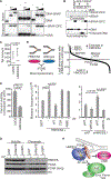
Figure 1.. HMCES is a ssDNA binding, replication fork associated protein.
(A) iPOND-SILAC-mass spectrometry derived nascent DNA/bulk chromatin abundance ratios for selected proteins or complexes are depicted. Mean+/−SD, n=3 or 4 for each cell line. (B) Immunoblot of wild type and HMCESΔ U2OS cells. (C) Clonogenic survival assay of cells treated with ATR inhibitor VX-970 for 24 hours. Mean+/−SD, n=3, ANOVA with Dunnett posttest. (D) Schematic diagram of HMCES and E. coli yedK. See also Figure S1. (E) Surface charge and catalytic pocket of the SRAP domain from human HMCES (PDB: 5KO9). (F-I) Electrophoretic mobility shift analysis of the indicated DNA ligands incubated with (F) GST-HMCES, (G) yedK-HIS, (H) wild type and mutant HMCES proteins after removal of the GST tag, or (I) wild type and mutant yedK-HIS proteins. See also Figure S2, Table S3, and Table S4.
Figure 2.
Figure 2.. HMCES localizes to chromatin in S-phase cells exposed to DNA damage and interacts with PCNA.
(A-B) RPE-hTERT cells that were contact inhibited (G0) then released for 20 hours to synchronize in S-phase (S) were compared to asynchronously growing cells. Immunoblots of (A) total cell lysates or (B) chromatin and soluble fractions. Cells were treated with 100 J/m2 UV and allowed to recover for 3h where indicated. (C) Flag-HMCES interacting proteins identified by mass spectrometry. Number of peptides identified for each protein in three experiments is indicated. Control cells do not express any tagged protein. (D) Proximity ligation assay with PCNA and HMCES antibodies. Scale bar is 10 μm. (E) Sequence alignment of the PIP box in HMCES compared to TLS polymerases. (F) Schematic diagram of HMCES indicating the location of the PIP box and the truncation mutants tested in G and H. (G) Flag-HMCES immunoprecipitates immunoblotted with Flag or PCNA antibodies. (H) Purified HMCES proteins were separated by SDS-PAGE and transferred to nitrocellulose. Membranes were incubated with purified PCNA and anti-PCNA antibodies.
Figure 3.
Figure 3.. HMCESΔ cells are hypersensitive to radiation and MMS.
(A) Clonogenic survival assay of wild type and HMCESΔ U2OS cells or siRNA-transfected cells treated with IR, UV, or MMS (siNT=non-targeting siRNA). (B) Immunoblot and (C) clonogenic survival analyses of HMCESΔ cells complemented with wild type and mutant HMCES. (D) IR sensitivity of wild type, HMCESΔ or the R98E mutation engineered into the HMCES genomic locus. (E) PARP inhibitor (BMN673) sensitivity of wild-type and HMCESΔ cells. All graphs show mean+/−SD, n=3, ANOVA with Dunnett post-test. See also Figures S3, S4, and Table S1.
Figure 4.
Figure 4.. HMCES crosslinks to AP sites and promotes AP site repair.
(A and B) Measurements of AP sites in genomic DNA using aldehyde reactive probe. All values are normalized to the untreated U2OS cell control. (A) Untreated U2OS versus HMCESΔ cells. mean+/−SD, n=8, one sample t-test, mean is different than 1. (B) Cells were treated with 10mM MMS (1 hour) and then allowed to recover for 1, 2 or 4 hours. Mean +/− SEM, n=5, ANOVA. (C) Denaturing gel analysis of 1nM unmodified (dT), THF stabilized abasic site, or a natural abasic site (dU + UDG) ssDNA incubated with human SRAP domain or yedK (3, 10nM). (D) Native and denaturing gel analysis of 1nM dU containing ssDNA or dsDNA treated with UDG as indicated. (E) Denaturing gel analysis of 1nM abasic site ssDNA (dU + UDG) incubated with wild type or mutant human SRAP (10, 1nM) or yedK (30, 10nM). Where indicated, proteinase K was used to digest the SRAP-DPC. (F) Measurement of abasic site DPC forming ability of GSTSRAP, *SRAP generated by cleavage of the GST leaving 4 N-terminal amino acids, and SRAPHIS tagged at the C-terminus. Mean+/−SD, n=3. (G) RADAR DPC assay: Mock or UV (100 J/m2) irradiated cells were allowed to recover for 3 hours in the presence or absence of MG132. Samples were lysed in detergents under denaturing conditions and DNA was ethanol precipitated. Only covalently attached proteins copurify with the DNA. 10μg of purified genomic DNA was digested with nuclease, applied to a nitrocellulose membrane, and immunoblotted with HMCES or RPA32 antibodies. (H) Quantification of RADAR assay on HMCESΔ cells complemented with wild type or mutant HMCES. Mean+/−SD, n=3, ANOVA with Dunnett posttest. See also Figures S2, S5, S6, and Table S3 and Table S4.
Figure 5.
Figure 5.. The HMCES-DPC is resolved by ubiquitin-dependent proteolysis
(A) Clonogenic survival assay of cells treated with KBrO3, mean+/−SD, N=3. Error bars in some cases are smaller than the symbols. (B) RADAR DPC assay: Cells were treated with 30mM KBrO3 for 30 minutes and allowed to recover for the indicated times in the presence or absence of MG132 (+). 20μg of purified genomic DNA was immunoblotted for HMCES. DNA was stained with methylene blue. (C) Quantification of B. Mean+/−SEM, n=3, two-tailed t-test. (D) Immunoblot analysis of total HMCES from cells treated with 30mM KBrO3 or 100 J/m2 UV. (E) Cells treated with 100 J/m2 UV were allowed to recover for 3 hours in the presence or absence of MG132, processed using the RADAR assay method. 100μg of purified genomic DNA was digested with nuclease, separated by SDS-PAGE, and immunoblotted for HMCES. (F) Cells expressing HMCES and HIS-ubiquitin were treated with 100 J/m2 UV and allowed to recover for 3 hours in the presence of MG132. HIS-tagged proteins purified under denaturing conditions were immunoblotted for HMCES. (G) Immunoblot analysis of total HMCES protein from cells treated with 100 J/m2 UV and incubated with or without MG132.
Figure 6.
Figure 6.. HMCES functions at replication forks to promote error-free repair of AP sites.
(A) Native and denaturing gel analysis of the indicated DNA ligands (1nM) treated with UDG to create an AP site after incubation with human HMCES SRAP domain (0, 1, or 10nM). (B) AP site containing ssDNA was incubated with SRAP domain and APE1 in the indicated order. Samples were resolved by SDS-PAGE and stained with coomassie or resolved on an 8M UREATBE gel prior to autoradiography. (C) DSBs were measured by neutral comet assay in wild type or HMCESΔ cells. Box and whiskers plot showing the median value, 25th to 75th percentile, and smallest and largest values; two-tailed t-test. (D) iPOND-SILAC-mass spectrometry was used to characterize the replication fork proteome of HMCESΔ compared to wild type HEK293T cells. Mean ratios, n=2. Two outlier data points were excluded from the graph but are included in Table S2. (E) Wild type or HMCESΔ cells were transfected with siRNA to REV3. The number of surviving colonies was compared to the non-targeting siRNA control in each cell line. Mean+/−SD, n=3, two-tailed t-test. (F) The mutation frequency of the indicated cell lines was assessed 48 hours after transfection of a mock or UV irradiated plasmid (pSP189), mean+/−SD, n=2, >50,000 colonies per sample, ANOVA with Dunnett post-test. (G) Cells were transfected with the indicated siRNAs. Immunoblot of chromatin and soluble fractions 3 hours after treating S-phase cells with 100 J/m2 UV. (H) Model of HMCES function. See also Figures S4, S6, and Tables S2–4.

References

    1. Amunugama R, Willcox S, Wu RA, Abdullah UB, El-Sagheer AH, Brown T, McHugh PJ, Griffith JD, and Walter JC (2018). Replication Fork Reversal during DNA Interstrand Crosslink Repair Requires CMG Unloading. Cell reports 23, 3419–3428. - PMC - PubMed
    1. Aravind L, Anand S, and Iyer LM (2013). Novel autoproteolytic and DNA-damage sensing components in the bacterial SOS response and oxidized methylcytosine-induced eukaryotic DNA demethylation systems. Biology direct 8, 20. - PMC - PubMed
    1. Bennardo N, Cheng A, Huang N, and Stark JM (2008). Alternative-NHEJ is a mechanistically distinct pathway of mammalian chromosome break repair. PLoS genetics 4, e1000110. - PMC - PubMed
    1. Bewick AJ, Vogel KJ, Moore AJ, and Schmitz RJ (2017). Evolution of DNA Methylation across Insects. Molecular biology and evolution 34, 654–665. - PMC - PubMed
    1. Bhat KP, and Cortez D (2018). RPA and RAD51: fork reversal, fork protection, and genome stability. Nat Struct Mol Biol 25, 446–453. - PMC - PubMed

Publication types

MeSH terms