. 2019 Jan 17;73(2):238-249.e3.
doi: 10.1016/j.molcel.2018.10.046. Epub 2018 Dec 13.

RSC-Associated Subnucleosomes Define MNase-Sensitive Promoters in Yeast

Affiliations

RSC-Associated Subnucleosomes Define MNase-Sensitive Promoters in Yeast

Sandipan Brahma et al. Mol Cell. .

Abstract

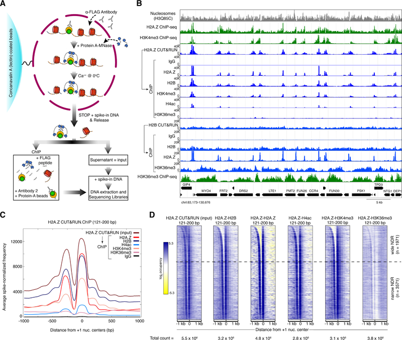
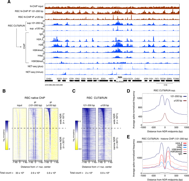
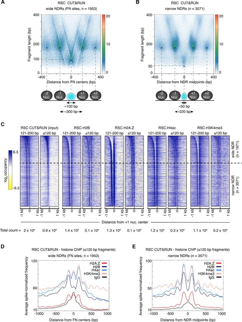
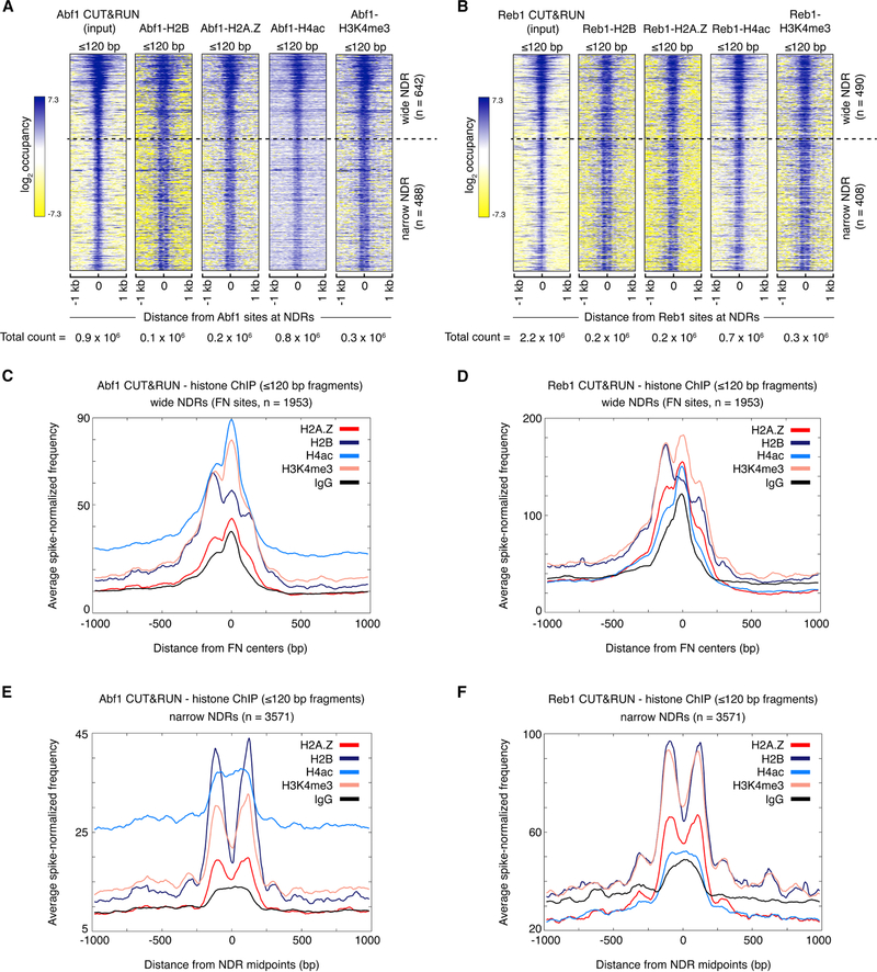
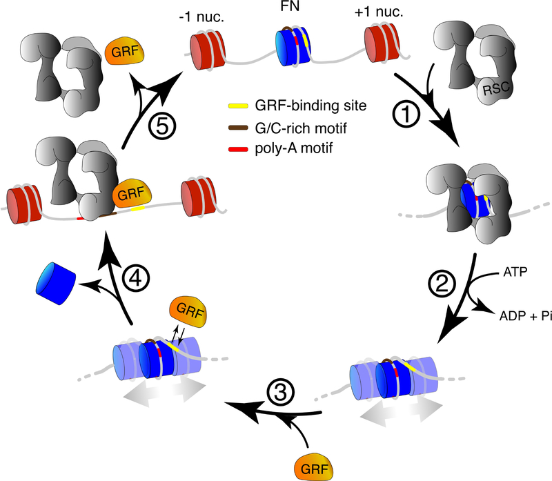
The classic view of nucleosome organization at active promoters is that two well-positioned nucleosomes flank a nucleosome-depleted region (NDR). However, this view has been recently disputed by contradictory reports as to whether wider (≳150 bp) NDRs instead contain unstable, micrococcal nuclease-sensitive ("fragile") nucleosomal particles. To determine the composition of fragile particles, we introduce CUT&RUN.ChIP, in which targeted nuclease cleavage and release is followed by chromatin immunoprecipitation. We find that fragile particles represent the occupancy of the RSC (remodeling the structure of chromatin) nucleosome remodeling complex and RSC-bound, partially unwrapped nucleosomal intermediates. We also find that general regulatory factors (GRFs) bind to partially unwrapped nucleosomes at these promoters. We propose that RSC binding and its action cause nucleosomes to unravel, facilitate subsequent binding of GRFs, and constitute a dynamic cycle of nucleosome deposition and clearance at the subset of wide Pol II promoter NDRs.

Keywords: CUT&RUN; fragile nucleosome; general regulatory factor; nucleosome dynamics; nucleosome-depleted region.


Conflict of interest statement

Declaration of interest

The authors declare no competing interests.

Figures

Figure 1:
Figure 1:. CUT&RUN.ChIP maps histone co-occupancies in single nucleosomes.
(A) Schematic diagram of the CUT&RUN.ChIP strategy. Yeast nuclei are successively treated with factor (or tag)-specific antibody (here anti-FLAG) and Protein A-MNase that diffuse into the nucleus. Ca++ induces MNase cleavage, and particles cleaved on both sides are released and diffuse out of the nucleus. FLAG-antibody is competed off from the released particles and antibody for a second epitope is added for ChIP followed by Protein-A beads to isolate immunoprecipitated particles. DNA extracted from the CUT&RUN supernatant and ChIP is used to prepare libraries for paired-end sequencing. Heterologous spike-in DNA can be added either before or after ChIP. (B) Spike-normalized (using the second spike-in DNA) CUT&RUN.ChIP profiles (121–200 bp) of histone modifications in H2A.Z and H2B nucleosomes (blue tracks). In each set (H2A.Z and H2B), the CUT&RUN supernatant was used as input for the subsequent ChIP-seq. IgG is a non-specific negative control for ChIP. The “Nucleosomes” track represents nucleosomal reads derived from H3Q85C chemical cleavage mapping of single-nucleosome dyads (Chereji et al., 2018). Crosslinking ChIP-seq of H2A.Z and the specified histone modifications (green tracks) are shown for comparison (Weiner et al., 2015). Tracks in each set of CUT&RUN.ChIP are scaled to the same intensity (y-axes). The positions of the annotated yeast open reading frames are shown at the bottom with the gene names. (C) Enrichment of spike-in normalized densities of 121–200 bp (nucleosomal) fragments from H2A.Z CUT&RUN and histone ChIP-seq using H2A.Z CUT&RUN as input, over the ±1-kb region spanning the dyads of 5542 +1 nucleosomes identified by H3Q85C cleavage mapping (Chereji et al., 2018). (D) Heat maps of 121–200 bp (nucleosomal) fragments from H2A.Z CUT&RUN.ChIP spanning +1 nucleosome centers ±1 kb, and sorted by NDR width. Heat maps are total count-normalized (total number of reads for each sample and size class are indicated at the bottom of each heat map), and shown in log2 scale, with log2 center = 5 and contrast = 3. Dotted lines separate wide NDRs (≥150-bp) from the rest. See also Figure S1.
Figure 2:
Figure 2:. CUT&RUN.ChIP maps and characterizes RSC-bound nucleosomes.
(A) Spike-normalized (using the second spike-in DNA) RSC CUT&RUN.ChIP profiles (blue) compared with RSC Native-ChIP (green) (Ramachandran et al., 2015). Reads of 121- to 200-bp were analyzed for the RSC CUT&RUN.ChIP profiles, where fragment sizes analyzed for the other tracks are indicated. NET-seq shows the relative number of reads at each nucleotide position corresponding to the 3′ ends of nascent RNA transcripts mapped to the yeast genome (Churchman and Weissman, 2011). Plus and minus denote reads mapped to the Watson and Crick strands, respectively. Yeast open reading frames are shown at the bottom in black, and a non-protein-coding RNA is shown in grey. (B, C) Heat maps of RSC Native-ChIP (B) and CUT&RUN (C) over 2-kb regions spanning the centers of the +1 nucleosomes, sorted by NDR width, and separated into 121–200 bp (nucleosomal) and ≤120 bp (subnucleosomal or short) size classes. Dotted lines separate wide NDRs (≥150-bp) from narrow NDRs. Heat maps are total count-normalized and shown in log2 scale, with log center = 1 and contrast = 2 (B, Native-ChIP) and log center = 5 and contrast = 4 (C, CUT&RUN). (D) Enrichment of spike-in normalized densities of ≤120 bp (subnucleosomal) and 121–200 bp (nucleosomal) fragments from RSC CUT&RUN.ChIP over the ±1-kb region spanning the midpoints of all annotated NDRs (Chereji et al., 2018). (E) Enrichment of spike-in normalized densities of 121–200 bp (nucleosomal) fragments from histone ChIP-seq of RSC CUT&RUN, over the ±1-kb region spanning the midpoints of all annotated NDRs. See also Figures S2 and S3.
Figure 3:
Figure 3:. RSC occupies wide NDRs and is associated with fragile nucleosomes.
(A, B) V-plots of RSC CUT&RUN spanning ±400 bp from annotated fragile nucleosome (FN) centers (Kubik et al., 2015) at ≳150 bp-wide NDRs (A) or the midpoints of narrow (all other) NDRs (B). Arrows show the positions corresponding to nucleosomal fragment midpoints, a subnucleosomal particle at a wide NDR (A), and a small particle over a narrow NDR (B) map. Horizontal dotted lines show the length in base pairs of limit CUT&RUN digestion around the central protected region in both plots. The average center-to-center distances between the +1 and −1 nucleosomes in both sets are shown at the bottom. For detailed interpretation of V-plots see Figure S3A. (C) Heat maps of RSC CUT&RUN.ChIP spanning +1 nucleosome centers ±1 kb, sorted by NDR width, and separated into 121–200 bp (nucleosomal) and ≤120 bp (subnucleosomal) size classes. Heat maps are total count-normalized (total number of reads for each sample and size class are indicated at the bottom of each heat map), and shown in log2 scale, with log center = 5 and contrast = 4. Dotted lines separate wide NDRs (≥150-bp) from narrow NDRs. (D, E) Enrichment of spike-in normalized densities of ≤120 bp fragments from RSC CUT&RUN.ChIP over the ±1-kb region spanning the NDRs, plotted separately for ≳150 bp-wide NDRs containing FN sites (D) and narrow (all other) NDRs (E). See also Figure S3.
Figure 4:
Figure 4:. GRFs associate with subnucleosomes at wide NDRs.
(A, B) Heat maps of ≤120 bp (subnucleosomal) fragments from Abf1 (A) and Reb1 (B) CUT&RUN.ChIP spanning ±1 kb of the respective GRF binding sites. Heat maps are total count-normalized and shown in log2 scale, with log center = 5 and contrast = 5. Dotted lines separate wide NDRs (≥150-bp) from the rest. Abf1 and Reb1 binding site locations were previously published (Skene and Henikoff, 2017). (C-F) Enrichment of spike-in normalized densities of ≤120 bp fragments from Abf1 (C, E) and Reb1 (D, F) CUT&RUN.ChIP over the ±1-kb region spanning the NDRs, plotted separately for ≳150 bp-wide NDRs containing fragile nucleosome (FN) sites (C, D) and narrow (all other) NDRs (E, F). See also Figures S4 and S5.
Figure 5:
Figure 5:. A dynamic model for RSC-mediated promoter nucleosome clearance and generation of fragile nucleosomes.
RSC, perhaps oriented by poly-A and G/C-rich sequence motifs, engulfs a nucleosome deposited at a 150 bp-wide NDR (step 1), and mobilizes and partially unwraps the nucleosome in an ATP-dependent manner (step 2). GRF binding is facilitated at sites exposed due to nucleosome displacement/unwrapping (step 3). RSC is still associated with the remodeled nucleosome at the two intermediate stages flanking step 3, but for simplicity RSC is not shown. RSC (and GRFs) eventually disrupt the nucleosome (step leading to stable GRF binding at sites on the exposed DNA. RSC may be also transiently bound to free DNA at the exposed NDRs, but not necessarily along with a GRF at the same time. When GRFs (and RSC) turn over (step 5), a nucleosome can reform at the NDR, and the cycle resumes.

References

    1. Badis G, Chan ET, van Bakel H, Pena-Castillo L, Tillo D, Tsui K, Carlson CD, Gossett AJ, Hasinoff MJ, Warren CL, et al. (2008). A library of yeast transcription factor motifs reveals a widespread function for Rsc3 in targeting nucleosome exclusion at promoters. Molecular cell 32, 878–887. - PMC - PubMed
    1. Bernstein BE, Mikkelsen TS, Xie X, Kamal M, Huebert DJ, Cuff J, Fry B, Meissner A, Wernig M, Plath K, et al. (2006). A bivalent chromatin structure marks key developmental genes in embryonic stem cells. Cell 125, 315–326. - PubMed
    1. Boeger H, Griesenbeck J, Strattan JS, and Kornberg RD (2003). Nucleosomes unfold completely at a transcriptionally active promoter. Molecular cell 11, 1587–1598. - PubMed
    1. Carey M, Li B, and Workman JL (2006). RSC exploits histone acetylation to abrogate the nucleosomal block to RNA polymerase II elongation. Molecular cell 24, 481–487. - PMC - PubMed
    1. Carrozza MJ, Li B, Florens L, Suganuma T, Swanson SK, Lee KK, Shia WJ, Anderson S, Yates J, Washburn MP, et al. (2005). Histone H3 methylation by Set2 directs deacetylation of coding regions by Rpd3S to suppress spurious intragenic transcription. Cell 123, 581–592. - PubMed

Publication types

MeSH terms