. 2018:51:e91.
doi: 10.1017/S0033583518000070. Epub 2018 Aug 3.

Key role of the REC lobe during CRISPR-Cas9 activation by 'sensing', 'regulating', and 'locking' the catalytic HNH domain

Affiliations

Key role of the REC lobe during CRISPR-Cas9 activation by 'sensing', 'regulating', and 'locking' the catalytic HNH domain

Giulia Palermo et al. Q Rev Biophys. 2018.

Abstract

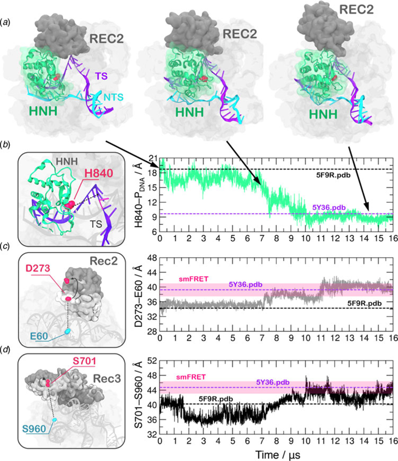
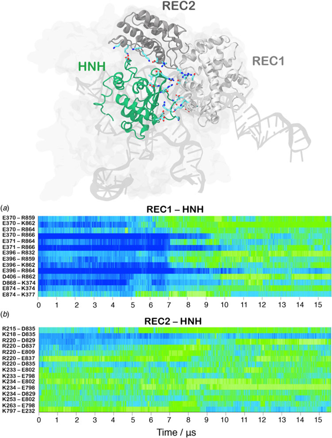
Understanding the conformational dynamics of CRISPR (clustered regularly interspaced short palindromic repeat)-Cas9 is of the utmost importance for improving its genome editing capability. Here, molecular dynamics simulations performed using Anton-2 - a specialized supercomputer capturing micro-to-millisecond biophysical events in real time and at atomic-level resolution - reveal the activation process of the endonuclease Cas9 toward DNA cleavage. Over the unbiased simulation, we observe that the spontaneous approach of the catalytic domain HNH to the DNA cleavage site is accompanied by a remarkable structural remodeling of the recognition (REC) lobe, which exerts a key role for DNA cleavage. Specifically, the significant conformational changes and the collective conformational dynamics of the REC lobe indicate a mechanism by which the REC1-3 regions 'sense' nucleic acids, 'regulate' the HNH conformational transition, and ultimately 'lock' the HNH domain at the cleavage site, contributing to its catalytic competence. By integrating additional independent simulations and existing experimental data, we provide a solid validation of the activated HNH conformation, which had been so far poorly characterized, and we deliver a comprehensive understanding of the role of REC1-3 in the activation process. Considering the importance of the REC lobe in the specificity of Cas9, this study poses the basis for fully understanding how the REC components control the cleavage of off-target sequences, laying the foundation for future engineering efforts toward improved genome editing.

Keywords: CRISPR–Cas9; genome editing; molecular dynamics; protein/nucleic acid interactions.


Conflict of interest statement

Conflict of interest. None.

Figures

Fig. 1.
Fig. 1.
(a) X-ray structure of the Streptococcus pyogenes CRISPR-Cas9 system (5F9R.pdb) in the pre-activated state (Jiang et al., 2016). Cas9 is shown in molecular surface, highlighting protein domains in different colors. The RNA (orange), the target DNA (TS, magenta), and non-target DNA (NTS, cyan) strands are shown as ribbons. (b) The catalytic residue H840 (magenta) of the HNH domain is located ~19 Å from the cleavage site on the TS. A close-up view of the active site shows the additional conformational change needed to attain the formation of an activated state (shown using an arrow).
Fig. 2.
Fig. 2.
(a) Conformational change of the HNH domain and structural adaptation of REC2 during ~16 µs of continuous MD simulations. (b–d) Time evolution along MD of: (b) the distance between H840 and the scissile phosphate on the target DNA strand (phosphate at position −3), indicating the approach of the HNH domain at the cleavage site; (c) the distance between atoms of E60 and D273 indicating the outward translation of REC2; (d) the distance between atoms of S960 and S701 indicating the conformational change of REC3. Horizontal bars are used to indicate the value of the three distances in the X-ray structure of the pre-activated state (5F9R.pdb, starting configuration for MD) and in the model structure obtained via cryo-EM fitting of the EMD-8236 map (5Y36.pdb) (Huai et al., 2017; Jiang et al., 2016). Transparent boxes are used to indicate the range assumed by the distances in single molecule Förster Resonance Energy Transfer (smFRET) experiments (Chen et al., 2017; Dagdas et al., 2017).
Fig. 3.
Fig. 3.
Time evolution along ~16 µs of continuous MD simulations of the CRISPR-Cas9 system of the salt-bridge interactions established between the HNH and REC1 domains (a) and between the HNH and REC2 domains (b). Salt bridges are computed as distance between the center of mass of the oxygen atoms in the acidic side chain and center of mass of the nitrogen atoms in the basic side chain. The scale on the right shows the change in strength of salt-bridge interactions: from weak (blue) to strong (green). The most important ionic interactions established by HNH and the REC1/REC2 regions are also shown on the three-dimensional structure of the activated CRISPR–Cas9.
Fig. 4.
Fig. 4.
Interaction established by the K810 (a), K848 (b), and K913 (c) residues of the HNH domain at ~0, 5, 10, 15 µs of MD. The RNA (gray), target DNA (TS, magenta), and non-target DNA (NTS, cyan) strands are shown as ribbons. The HNH domain (green) is shown as cartoon. Interacting residues are shown as sticks.
Fig. 5.
Fig. 5.
(a) Matrix of the generalized correlation (GC), calculated over the last ~4 µs of MD of CRISPR–Cas9. The strength of the computed correlations is color-coded from green (highly correlated motions) to gray (poorly correlated motions). A box is used to highlight the high correlation between the HNH domain and the REC lobe. (b) Inter-domain GC scores (GCs) plotted for each Cas9 domain (REC1–3, HNH, RuvC, and PI domain) in a two-by-two matrix, measuring the strength of the overall correlation that each domain establishes with the others (details in the Method section). (c) The highest GCs are plotted on the three-dimensional structure of CRISPR–Cas9, revealing a tight coupling between HNH and the REC2–3 domains. Cas9 is shown in a molecular surface, highlighting the HNH and REC2–3 domains with different colors. The RNA (orange) and the target DNA (TS, magenta) forming a RNA:DNA hybrid, as well as the non-target DNA (NTS, cyan) are shown as ribbons.

References

    1. Abrams C and Bussi G (2014) Enhanced sampling in molecular dynamics using metadynamics, replica-exchange, and temperature-acceleration. Entropy 16, 163–199.
    1. Amadei A, Linssen ABM and Berendsen HJC (1993) Essential dynamics of proteins. Proteins-Structure Function and Genetics 17, 412–425. - PubMed
    1. Anders C, Niewoehner O, Duerst A and Jinek M (2014) Structural basis of PAM-dependent target DNA recognition by the Cas9 endonuclease. Nature 513, 569–573. - PMC - PubMed
    1. Aqvist J (1990) Ion-water interaction potentials derived from free energy per-turbation simulations. Journal of Physical Chemistry 94, 8021–8024.
    1. Banas P, Hollas D, Zgarbova M, Jurecka P, Orozco M, Cheatham TE, Sponer J and Otyepka M (2010) Performance of molecular mechanics force fields for RNA simulations: stability of UUCG and GNRA hairpins. Journal of Chemical Theory and Computation 6, 3836–3849. - PMC - PubMed

Publication types

Substances