. 2018 Nov 28:9:1368.
doi: 10.3389/fphar.2018.01368. eCollection 2018.

IFN-β Plays Both Pro- and Anti-inflammatory Roles in the Rat Cardiac Fibroblast Through Differential STAT Protein Activation

Affiliations

IFN-β Plays Both Pro- and Anti-inflammatory Roles in the Rat Cardiac Fibroblast Through Differential STAT Protein Activation

Samir Bolívar et al. Front Pharmacol. .

Abstract

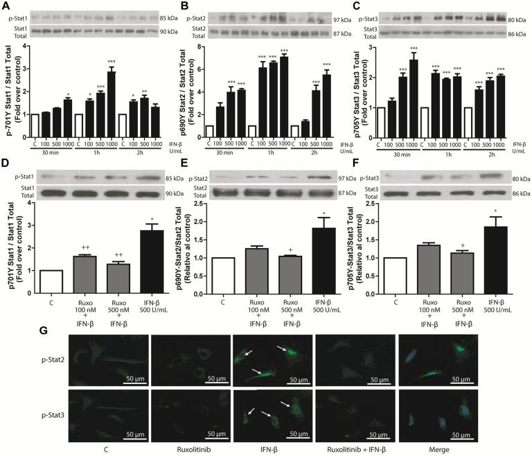
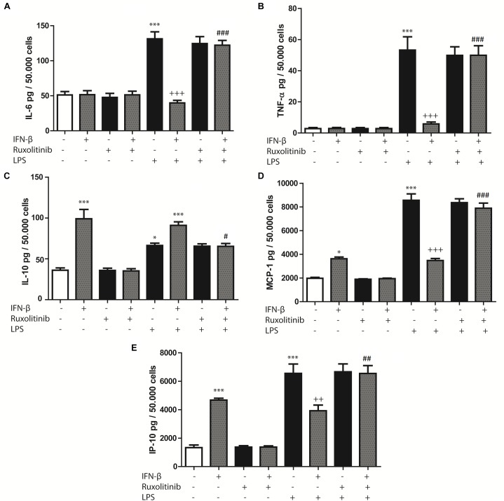
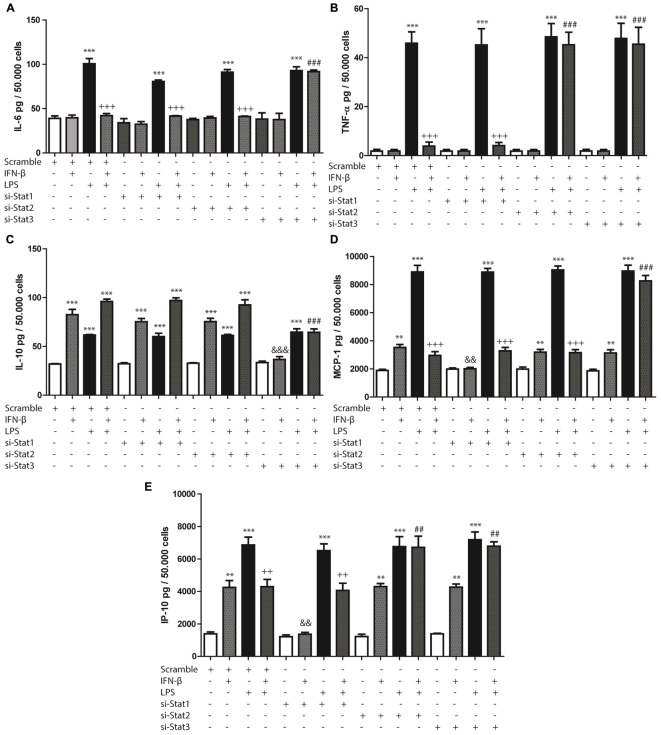
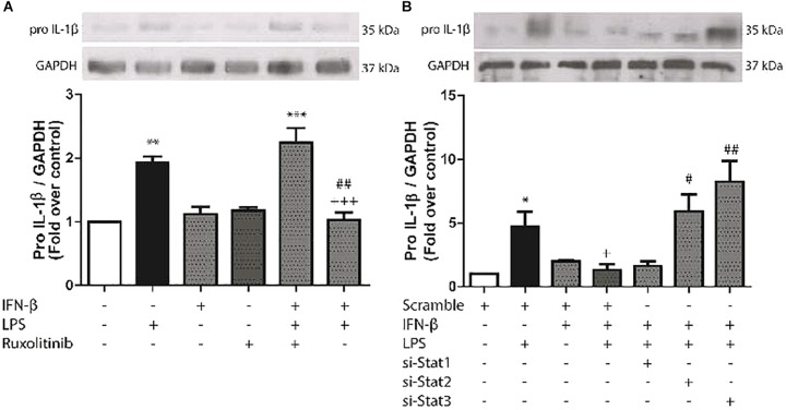
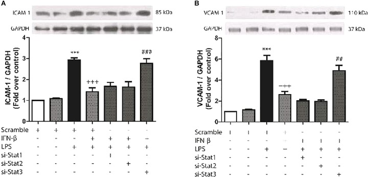
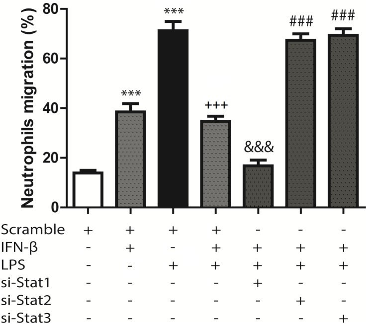
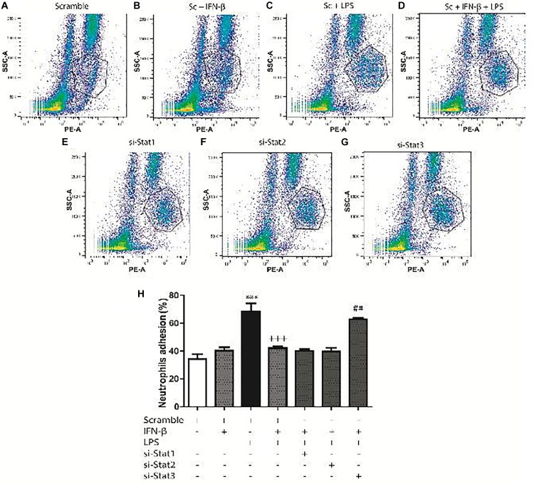
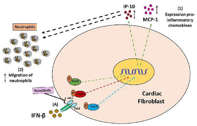
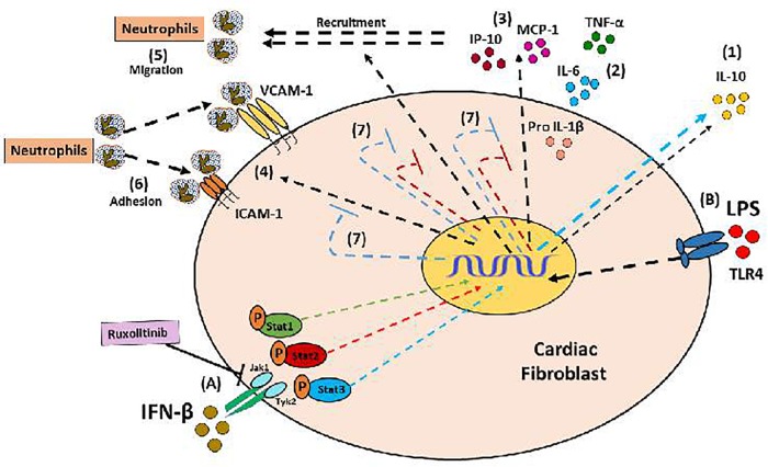
Cardiac fibroblasts (CFs) contribute to theinflammatory response to tissue damage, secreting both pro- and anti-inflammatory cytokines and chemokines. Interferon beta (IFN-β) induces the phosphorylation of signal transducer and activator of transcription (STAT) proteins through the activation of its own receptor, modulating the secretion of cytokines and chemokines which regulate inflammation. However, the role of IFN-β and STAT proteins in modulating the inflammatory response of CF remains unknown. CF were isolated from adult male rats and subsequently stimulated with IFN-β to evaluate the participation of STAT proteins in secreting chemokines, cytokines, cell adhesion proteins expression and in their capacity to recruit neutrophils. In addition, in CF in which the TRL4 receptor was pre-activated, the effect of INF-β on the aforementioned responses was also evaluated. Cardiac fibroblasts stimulation with IFN-β showed an increase in STAT1, STAT2, and STAT3 phosphorylation. IFN-β stimulation through STAT1 activation increased proinflammatory chemokines MCP-1 and IP-10 secretion, whereas IFN-β induced activation of STAT3 increased cytokine secretion of anti-inflammatory IL-10. Moreover, in TLR4-activated CF, IFN-β through STAT2 and/or STAT3, produced an anti-inflammatory effect, reducing pro-IL-1β, TNF-α, IL-6, MCP-1, and IP-10 secretion; and decreasing neutrophil recruitment by decreasing ICAM-1 and VCAM-1 expression. Altogether, our results indicate that IFN-β exerts both pro-inflammatory and anti-inflammatory effects in non-stimulated CF, through differential activation of STAT proteins. When CF were previously treated with an inflammatory agent such as TLR-4 activation, IFN-β effects were predominantly anti-inflammatory.

Keywords: IFN-β (interferon β); STAT; anti-infammatory; cardiac fibroblast; proinflammatory.


Figures

FIGURE 1
FIGURE 1
IFN-β activates the JAK/STAT signaling pathway in CF. (A–C) The upper panelshows a Western blot image, indicating the INF-β-induced p-STAT1, p-STAT2, and p-STAT3 expression levels; STAT1, STAT2, and STAT3 were used as loading controls. The graphical analysis is presented at the bottom of each panel. Error bars indicate theSD for four independent experiments. ∗∗∗p < 0.001, ∗∗p < 0.01, p < 0.05 vs. control. (D–F) CF were pretreated with or without Ruxolitinib (100 and 500 nM) for 1 h and stimulated with IFN-β (500 U/ml) for 24 h. The upper panel of each figure corresponds to the representative image of p-STAT1, p-STAT2, and p-STAT3 expression levels; STAT1, STAT2, and STAT3 were used as loading controls. The graphical analysis is presented at the bottom of each panel. Error bars indicate the SD for four independent experiments. ∗∗∗p < 0.001, p < 0.05 vs. control. ++p < 0.01, p < 0.05 vs. IFN-β. (G) CF were pretreated with or without Ruxolitinib (100 and 500 nM) for 1 h and stimulated with IFN-β (500 U/ml) for 24 h. p-STAT2 and p-STAT3 were detected by immunofluorescence using anti-p-STAT2 and anti-p-STAT3 antibodies and Alexa Fluor® 488-conjugated secondary antibody (green staining). Representative images of the immunocytochemistry for three independent experiments are shown, indicating the nuclear translocation of p-STAT2 and p-STAT3. +p < 0.05 vs. IFN-β.
FIGURE 2
FIGURE 2
Effects of IFN-β and STAT proteins on LPS-induced cytokine secretion: (A–E) CF were transfected with scramble or 200 ng of si-STAT1, si-STAT2, and si-STAT3 for 8 h, serum-deprived of for 24 h, pre-treated with IFN-β for 1 h, and stimulated with LPS for 24 h. (A) IL-6, (B) TNF-β, (C) IL-10, (D) MCP-1 and (E) IP-10 secretion to the culture medium was determined by LUMINEX assay. Error bars indicate the SD for three independent experiments. ∗∗∗p < 0.001, ∗∗p < 0.01, vs. scramble. +++p < 0.001 vs. scramble + LPS. ###p < 0.001, ##p < 0.01 vs. scramble + IFN-β + LPS. &&&p < 0.001, &&p < 0.01 vs. scramble + IFN-β.
FIGURE 3
FIGURE 3
Effects of IFN-β and STAT proteins on LPS-induced cytokine secretion: (A–E) CF were transfected with scramble or 200 ng of si-STAT1, si-STAT2, and si-STAT3 for 8 h, serum-deprived of for 24 h, pre-treated with IFN-β for 1 h, and stimulated with LPS for 24 h. (A) IL-6, (B) TNF-β, (C) IL-10, (D) MCP-1 and (E) IP-10 secretion to the culture medium was determined by LUMINEX assay. Error bars indicate the SD for three independent experiments. ∗∗∗p < 0.001, ∗∗p < 0.01, vs. scramble. +++p < 0.001 vs. scramble + LPS. ###p < 0.001, ##p < 0.01 vs. scramble + IFN-β + LPS. &&&p < 0.001, &&pp < 0.01 vs. scramble + IFN-β.
FIGURE 4
FIGURE 4
Effects of IFN-β and STAT proteinson LPS-induced pro-IL-1β protein expression. (A) CF were pretreated with or without Ruxolitinib (500 nM) for 1 h and stimulated with IFN-β (500 U/ml) for 24 h. The upper panel shows a representative Western blot image, indicating expression levels of pro-IL-1β protein and GAPDH (used as charge control). The graphical analysis is presented at the bottom of the panel. Error bars indicate the SD for three independent experiments. ∗∗∗p < 0.001, ∗∗p < 0.01 vs. control. ##p < 0.01 vs. LPS. +++p < 0.001 vs. Ruxo + IFN-β + LPS). (B) CF were transfected with scramble or 200 ng of si-STAT1, si-STAT2, and si-STAT3 for 8 h, serum-deprived for 24 h, pretreated with IFN-β (500 U/ml) for 1 h, and stimulated with LPS (1 μg/ml) for 8 h. The upper panel shows a Western blot image, indicating expression levels of pro-IL-1β protein and GAPDH (used as charge control). The graphical analysis is presented at the bottom of the panel. Error bars indicate the SD for three independent experiments. p < 0.05 vs. scramble. +p < 0.05 vs. scramble + LPS. ##p < 0.01, #p < 0.05 vs. scramble + IFN-β + LPS.
FIGURE 5
FIGURE 5
IFN-β decreases LPS-induced ICAM-1/VCAM-1 expression through STAT3. (A–B) CF were transfected with scramble or 200 ng of si-STAT1, si-STAT2, and si-STAT3 for 8 h, serum-deprived for 24 h, pretreated with IFN-β (500 U/ml) for 1 h, and stimulated with LPS (1 μg/ml) for 8 h. (A) The upper panel shows a representative Western blot image, indicating expression levels of ICAM-1 and GAPDH (used as charge control). The graphical analysis is presented at the bottom of each panel. Error bars indicate the SD for three independent experiments. ∗∗∗p < 0.001 vs. scramble. +++p < 0.001 vs. scramble + LPS. ###p < 0.001 vs. scramble + IFN-β + LPS. (B) The upper panel shows a representative Western blot image, indicating expression levels of VCAM-1 and GAPDH (used as charge control). The graphical analysis is presented at the bottom of each panel. Error bars indicate the SD for three independent experiments. ∗∗∗p < 0.001 vs. scramble. +++p < 0.001 vs. scramble + LPS. ##p < 0.01 vs. scramble + IFN-β + LPS.
FIGURE 6
FIGURE 6
Neutrophil migration induced by conditioned culture media derived from IFN-β- and LPS-treated CF: assays were performed in transwell chambers. Neutrophils charged with fluorescent calcein were seeded in the upper chamber and allowed to migrate at 37°C for 3 h to the lower chamber containing conditioned culture medium derived from CF that were transfected with scramble or 200 ng of STAT1 siRNA, STAT2, and STAT3 for 8 h, serum-deprived for 24 h, pretreated with IFN-β (500 U/ml) for 1 h, and stimulated with LPS (1 μg/ml) for 8 h. Fluorescent neutrophils that migrated to the lower compartment were measured using a fluorescence spectrometer, and the chemotactic activity was expressed as the percentage of fluorescent cells in the lower chamber as compared to the fluorescence of the total neutrophils added. Error bars indicate the SD for three independent experiments. ∗∗∗p < 0.001 vs. scramble. +++p < 0.001 vs. scramble + LPS. &&&p < 0.001 vs. scramble + IFN-β. ###p < 0.001 vs. scramble + IFN-β + LPS.
FIGURE 7
FIGURE 7
IFN-β decreases neutrophil adhesion to CF. Leukocytes were labeled with anti-RP1/PE (neutrophil marker) and incubated for 2 h at 37°C with confluent monolayers of CF transfected with scramble or 200 ng si-STAT and treated with IFN-β and LPS. Cells were analyzed using flow cytometry. (A–G) FACS dots plots are representative images of the percentage of neutrophils adhered to CF. (H) Graphical analysis of the percentage of neutrophils in co-culture adhered to scramble-transfected CF with no treatment or treated with IFN-β or LPS. The neutrophil adhesion index was calculated was as follows: 100 x [no. cells (RP1 +) adhered/total number of cells (RP-1 +)]. Error bars indicate the SD for three independent experiments. ∗∗∗p < 0.001 vs. scramble. +++p < 0.001 vs. scramble + LPS. ##p < 0.01 vs. scramble + IFN-β + LPS.
FIGURE 8
FIGURE 8
Proinflammatory and anti-inflammatory effects of IFN-β in CF: (A) IFN-β triggers JAK/STATsignaling pathway activation (STAT1, STAT3, and STAT3 proteins). (1) IFN-β induces a pro-inflammatory response in the CF through STAT1 protein activation (green arrows), characterized by increased expression of the chemokines IP-10 and MCP-1, as well as (2) increased neutrophil migration, favored by chemokines released from CF.
FIGURE 9
FIGURE 9
Anti-inflammatory effect of IFN-β in CF: (A) IFN-β induces the activation of its JAK/STAT transduction pathway (proteins Stat1, Stat3, Stat3); and directly through Stat3 induces the secretion of IL-10 (1). (B) The LPS is a pro-inflammatory stimulus that favors the secretion of cytokines (2), chemokines (3), expression of adhesion molecules (4), which finally favors the recruitment of neutrophils by the FC (5 and 6) . (7) IFN-β through the activation of Stat2 and Stat3 proteins (red and blue STOP lines), inhibits the pro-inflammatory effects of LPS.

References

    1. Aas V., Lappegård K. T., Siebke E. M., Benestad H. B. (1996). Modulation by interferons of human neutrophilic granulocyte migration. J. Interferon Cytokine Res. 16 929–935. 10.1089/jir.1996.16.929 - DOI - PubMed
    1. Boza P., Ayala P., Vivar R., Humeres C., Tapia Cáceres F., Muñoz C., et al. (2016). Expression and function of toll-like receptor 4 and inflammasomes in cardiac fibroblasts and myofibroblasts: IL-1β synthesis, secretion, and degradation. Mol. Immunol. 74 96–105. 10.1016/j.molimm.2016.05.001 - DOI - PubMed
    1. Braunwald E., Kloner R. A. (1985). Myocardial reperfusion: a double-edged sword? J. Clin. Invest. 76 1713–1719. 10.1172/JCI112160 - DOI - PMC - PubMed
    1. Chen J., Baig E., Fis E. N. (2004). Diversity and relatedness among the type I interferons. J. Interferon Cytokine Res. 24 687–698. 10.1089/jir.2004.24.687 - DOI - PubMed
    1. Coclet-Ninin J., Dayer J. M., Burger D. (1997). Interferon-beta not only inhibits interleukin-1beta and tumor necrosis factor-alpha but stimulates interleukin-1 receptor antagonist production in human peripheral blood mononuclear cells. Eur. Cytokine Netw. 8 345–349. - PubMed