

Effect of Polymethylmethacrylate-Hydroxyapatite Composites on Callus Formation and Compressive Strength in Goat Vertebral Body
- PMID: 30555640
- PMCID: PMC6287135
- DOI: 10.5704/MOJ.1811.002
Effect of Polymethylmethacrylate-Hydroxyapatite Composites on Callus Formation and Compressive Strength in Goat Vertebral Body
Abstract
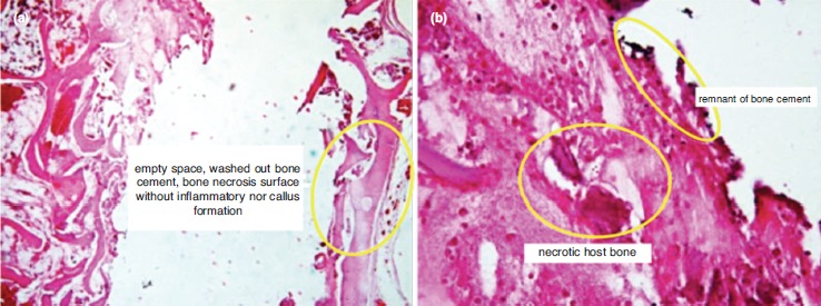
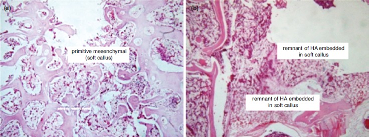
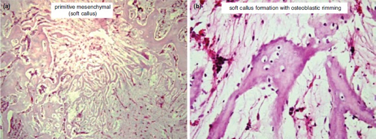
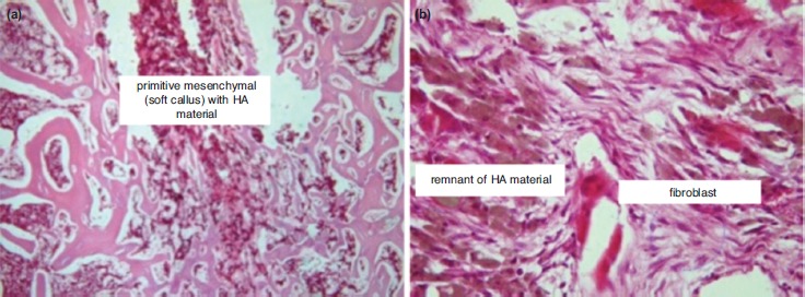
Introduction: Percutaneous vertebroplasty (PV) is one of the available treatments for vertebral compression fracture (VCF). Polymethylmethacrylate (PMMA) is the most common bone substitute used in the procedure, but it has several disadvantages. Bioceramic material, such as hydroxyapatite (HA), has better biological activity compared to PMMA. The aim of this study was to find an optimal biomaterial compound which offers the best mechanical and biological properties to be used in PV. Materials and Methods: This was an experimental study with goat (Capra aegagrus hircus) as an animal model. The animals' vertebral columns were injected with PMMA-HA compound. Animal samples were divided into four groups, and each group received a different proportion of PMMA:HA compound. The mechanical and biological effects of the compound on the bone were then analysed. The mechanical effect was assessed by measuring the vertebral body's compressive strength. Meanwhile, the biological effect was assessed by analysing the callus formation in the vertebral body. Results: The optimal callus formation and compressive strength was observed in the group receiving PMMA:HA with a 1:2 ratio. Conclusion: A mixture of PMMA and HA increases the quality of callus formation and the material's compressive strength. The optimum ratio of PMMA:HA in the compound is 1:2.
Keywords: hydroxyapatite; polymethylmethacrylate; vertebral compression fracture; vertebroplasty.
Figures
References
-
- Predey TA, Sewall LE, Smith SJ. Percutaneous vertebroplasty: new treatment for vertebral compression fractures. Am Fam Physician. 2002;66(4):611–5. - PubMed
-
- Teotia AK, Raina DB, Singh C, Sinha N, Isaksson H, Tägil M et al. Nano-hydroxyapatite bone substitute functionalized with bone active molecules for enhanced cranial bone regeneration. ACS Appl Mater Interfaces. 2017;9(8):6816–28. - PubMed
-
- Matsumine A, Myoui A, Kusuzaki K, Araki N, Seto M, Yoshikawa H et al. Calcium hydroxyapatite ceramic implants in bone tumour surgery. A long term follow-up study. J Bone Joint Surg Br. 2004;86(5):719–25. - PubMed
LinkOut - more resources
Full Text Sources