Review
. 2018 Nov 1;8(11):2165-2175.
eCollection 2018.

Exosomes in cancer therapy: a novel experimental strategy

Affiliations
Review

Exosomes in cancer therapy: a novel experimental strategy

Dong Gao et al. Am J Cancer Res. .

Abstract

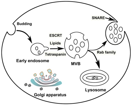
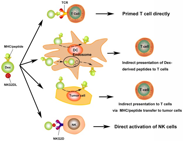
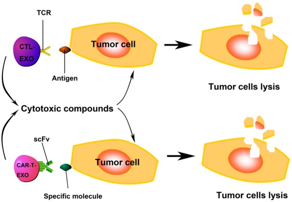
Exosomes are small membrane vesicles of endocytic origin secreted by most cell types. They play important roles in intercellular communications and many physiological processes. DCs-derived exosomes can prime naïve T cells and activate NK cells to shrink the tumor. Tumor-derived exosomes carry a variety of tumor antigens that trigger the robust tumor antigen-specific immune response. Tumor-derived exosomes also contain metastasis or invasive-related molecules, which maybe potential targets for cancer immunotherapy. Effector T cells-derived exosomes possess cytotoxic activity of their original cells, thus cause tumor cells lysis. In this review, we summarized the recent advances on the biogenesis and composition of exosomes, the functions of anti-tumor immune response, and the promising applications on cancer immunotherapy of exosomes from different origins. Exosomes schlep efficient targets homing to tumor sites and tend to be a promising new tool of immunotherapy to fight cancer in a cell-free system.

Keywords: CAR-T cells; CTLs; DCs; Exosomes; cancer immunotherapy.


Conflict of interest statement

None.

Figures

Figure 1
Figure 1
Schematic representation of the biogenesis and release of exosomes by eukaryotic cells. Exosomes firstly formed as multivesicular body (MVB) by budding into early endosomes. Several mechanisms including ESCRT, lipids and tetraspanin-dependent, are involved in MVB formation. If MVBs did not target for lysosomal degradation, they may mobilize and fuse with plasma membrane (PM) to release exosomes, depending on Rab family and soluble NSF-attachment protein receptor (SNARE) complexes respectively.
Figure 2
Figure 2
The role of exosomes derived from DC (Dexs) in anti-tumor immunity. Dexs may stimulate T cells via direct and indirect routes. The presence of MHC/peptide complex on the surface of Dexs stimulates T cells directly. Dexs activate T cells indirectly by bystander DCs via two mechanisms, one is Dexs fuse with PM of DCs and transfer MHC/peptide complexes to DCs surface immediately, the other one, Dexs are internalised into the endosomes and processed by APCs, following the antigen presentation on the surface of APCs indirectly. Dexs may also present MHC/peptide complexes to host T cells by MHC/peptide transfer to tumor cells. Dexs were shown to possess NKG2D ligand (NKG2DL), which can interact with NKG2D and activate NK cells.
Figure 3
Figure 3
The role of tumor cells secreted exosomes (Texs) in anti-tumor immunity. Texs expressed tumor MHC/antigen complexes internalised by or fused with antigen-presenting cells (APCs) to prime T cells. Stress-inducible exosomal HSP70 functions as an endogenous danger signal, promotes NK cell activation and cancer cell lysis via granzyme B. Texs improve the HCC tumor microenvironment in vivo after infusion.
Figure 4
Figure 4
The role of exosomes derived from CTLs (CTL-EXO) and CAR-T cells (CAR-T-EXO) in anti-tumor immunity. The presence of TCR/CD3/CD8 on CTL-EXO enable CTL-EXO target antigen expressing tumor cells specificly, and cytotoxic compounds contained in CTL-EXO kill the target cells. CAR-T-EXO may possess antibody-derived single-chain variable fragment (scFv), which determines the targeting specificity of CAR-T cells, can recognize specific molecules on target cells (such as CD19 on B-cell neoplasms) and kill them by cytotoxic compounds release.

Similar articles

Cited by

References

    1. Johnstone RM, Adam M, Hammond JR, Orr L, Turbide C. Vesicle formation during reticulocyte maturation. Association of plasma membrane activities with released vesicles (exosomes) J Biol Chem. 1987;262:9412–9420. - PubMed
    1. Pan BT, Teng K, Wu C, Adam M, Johnstone RM. Electron microscopic evidence for externalization of the transferrin receptor in vesicular form in sheep reticulocytes. J Cell Biol. 1985;101:942–948. - PMC - PubMed
    1. Harding C, Heuser J, Stahl P. Receptor-mediated endocytosis of transferrin and recycling of the transferrin receptor in rat reticulocytes. J Cell Biol. 1983;97:329–339. - PMC - PubMed
    1. Colombo M, Moita C, van Niel G, Kowal J, Vigneron J, Benaroch P, Manel N, Moita LF, Thery C, Raposo G. Analysis of ESCRT functions in exosome biogenesis, composition and secretion highlights the heterogeneity of extracellular vesicles. J Cell Sci. 2013;126:5553–5565. - PubMed
    1. Thery C, Ostrowski M, Segura E. Membrane vesicles as conveyors of immune responses. Nat Rev Immunol. 2009;9:581–593. - PubMed

LinkOut - more resources