

doi: 10.1016/j.ctro.2018.11.003.
eCollection 2019 Jan.
Visualization of the tumor cavity after lumpectomy of breast cancer for postoperative radiotherapy
Affiliations
- PMID: 30555941
- PMCID: PMC6279963
- DOI: 10.1016/j.ctro.2018.11.003
Item in Clipboard
Visualization of the tumor cavity after lumpectomy of breast cancer for postoperative radiotherapy
Clin Transl Radiat Oncol.
.
Abstract
To visualize the tumor cavity after lumpectomy, the tumor cavity was coated with the liquid tissue marker sucrose acetate isobutyrate (SAIB) with its radiopaque electron dense SAIB analogue (x-SAIB) and assessed for radiotherapy planning. SAIB/x-SAIB enhanced the confidence for target structure definition. Tissue displacement after oncoplasty may be revealed by SAIB/x-SAIB.
Figures
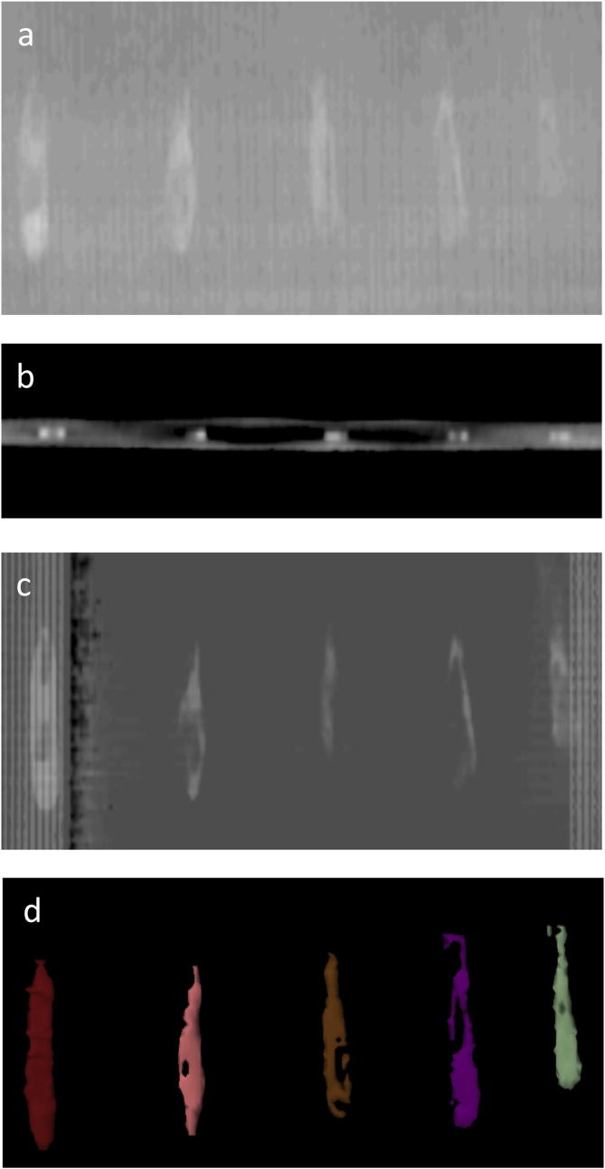
Assessment of visibility of SAIB/X-SAIB for radiotherapy planning on a phantom. Different amounts of SIAB/x-SIAB were applied (a) over a length of 5 cm on a paper carrier with the fingertip loaded with (from left to right) with 50 mikroL, 40 mikrolL, 30 mikroL, 20 mikroL or 10 mikroL. (a) shows a plain X-ray of the marker, (b) the axial imaging on a planning CT at a slice thickness of 3 mm, (c) a digitally reconstructed radiograph, and (d) segmentation of the liquid tissue marker.
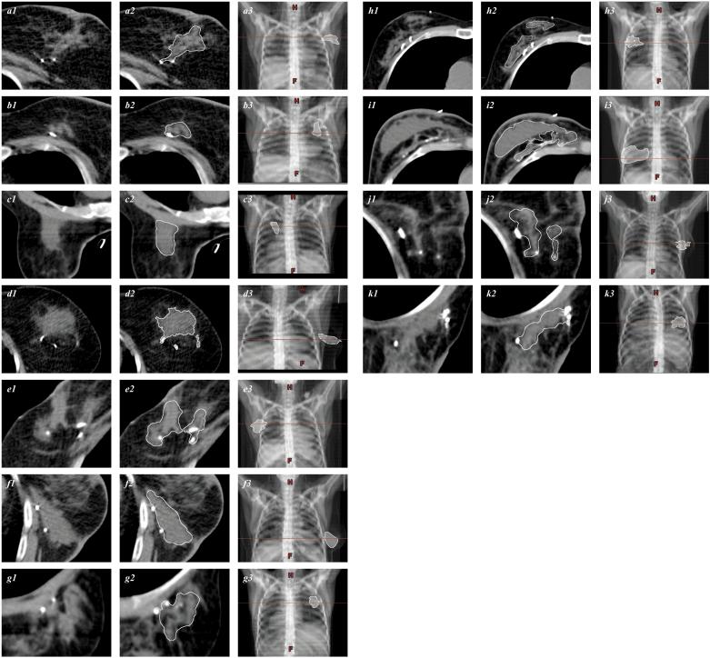
Contrast-enhanced visualization of the tumor cavity following lumpectomy with SIAB/x-SIAB. Scans shown are gated from (−132)–(−124) to (201)–(225) Hounsfield units. Patients underwent tumor excision and adjuvant EBRT (a to g), or tumor excision followed by oncoplastic surgery (h to k). Patients in c, g, and k were treated in prone position. Figures a1…k1 indicate the unprocessed imaging data. Manual segmentation target structure definition is shown in a2…k2 (white line). Figures a3…k3 show the digital reconstructed radiograph with the horizontal line passing trough the target structure (white) and the corresponding axial sections.
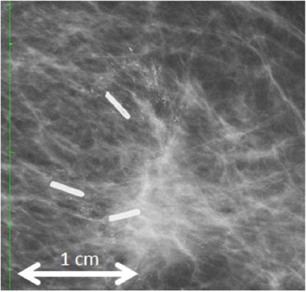
Follow-up X-ray imaging after tumorectomy. The green scale indicates 1 cm between ticks. One year after definitive surgery and adjuvant radiotherapy, a mammogram reveals granular residual radiopaque traces mimicking microcalcifications.
References
-
- Correa C., Harris E.E., Leonardi M.C. Accelerated partial breast irradiation: executive summary for the update of an astro evidence-based consensus statement. Pract Radiat Oncol. 2017;7:73–79. - PubMed
-
- Gonzalez Sanchis A., Brualla Gonzalez L., Fuster Diana C. Tumor bed segmentation: first step for partial breast irradiation. Clin Transl Oncol. 2013;15:39–45. - PubMed
LinkOut - more resources
Full Text Sources
