Review
. 2018 Jul 10;30(4):297-310.
doi: 10.1016/j.joco.2018.06.004. eCollection 2018 Dec.

Deep anterior lamellar keratoplasty: A surgeon's guide

Affiliations
Review

Deep anterior lamellar keratoplasty: A surgeon's guide

Mayank A Nanavaty et al. J Curr Ophthalmol. .

Abstract

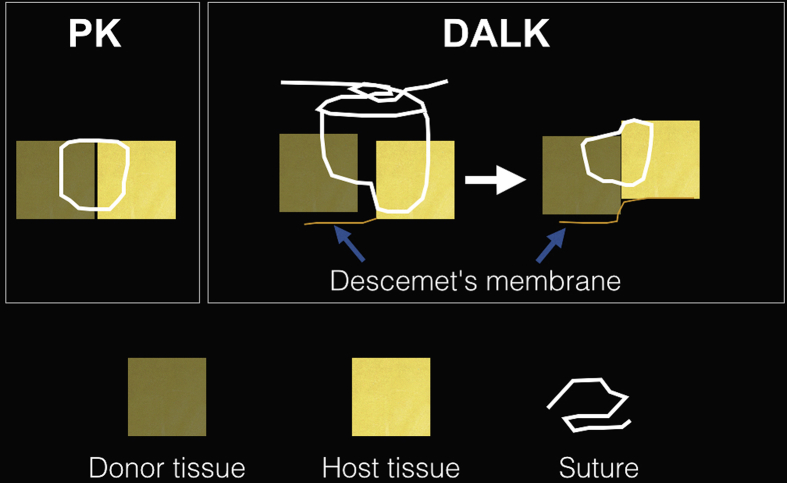
Purpose: To review and highlight important practical aspects of deep anterior lamellar keratoplasty (DALK) surgery and provide some useful tips for surgeons wishing to convert to this procedure from the conventional penetrating keratoplasty (PK) technique.

Methods: In this narrative review, the procedure of DALK is described in detail. Important pre, intra, and postoperative considerations are discussed with illustrative examples for better understanding. A comprehensive literature review was conducted in PubMed/Medline from January 1995 to July 2017 to identify original studies in English language regarding DALK. The primary endpoint of this review was the narrative description of surgical steps for DALK, its pitfalls, and management of common intraoperative complications.

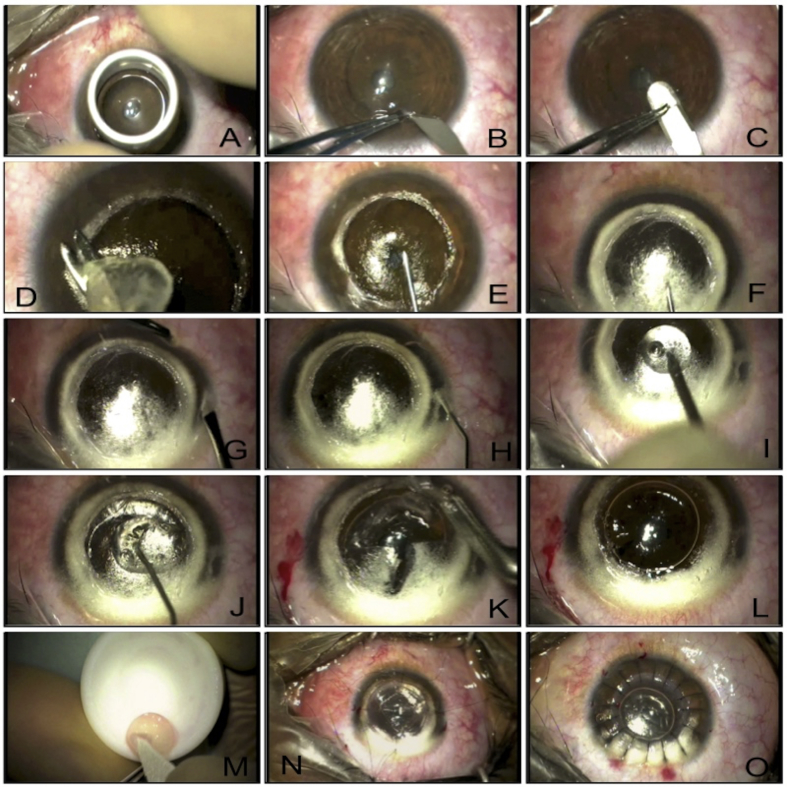
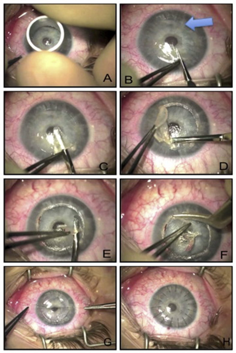
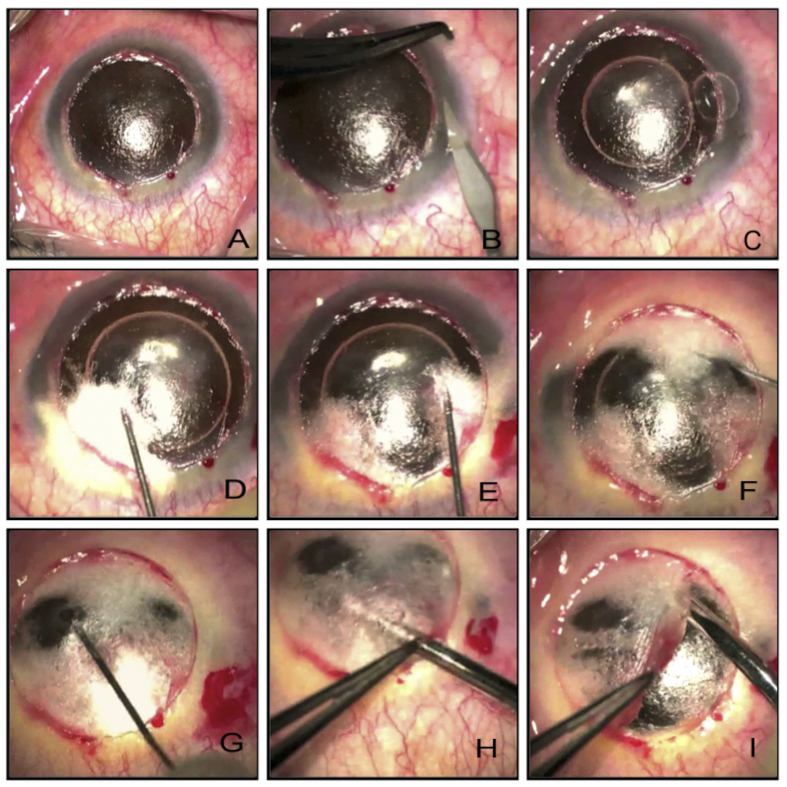
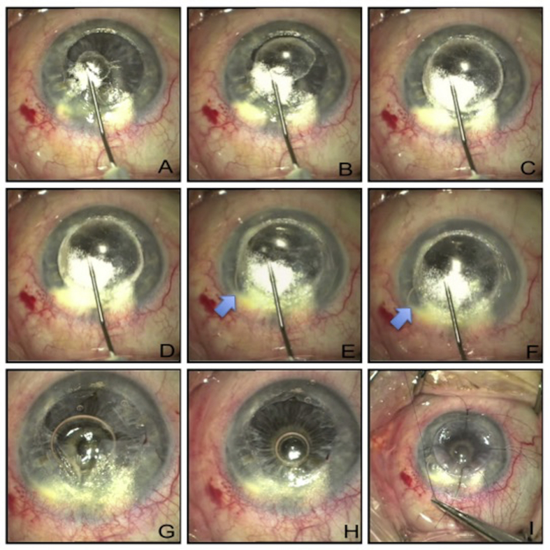
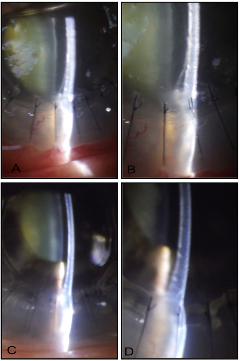
Results: A standard DALK procedure can be successfully performed taking into consideration factors such as age, ophthalmic co-morbidities, status of the crystalline lens, retina, and intraocular pressure. Careful trephination and dissection of the host cornea employing appropriate technique (such as big bubble technique, manual dissection, visco-dissection, etc.) suitable for the specific case is important to achieve good postoperative outcomes. Prompt identification of intraoperative complications such as double bubble, micro and macroperforations, etc. are vital to change the management strategies.

Conclusion: Although there is a steep learning curve for DALK procedure, considering details and having insight into the management of intraoperative issues facilitates learning and reduces complication rates.

Keywords: DALK; Keratoplasty; Lamellar corneal transplant.


Figures

Fig. 1
Fig. 1
Dehydration of the Teflon block – corneal tissue interface using a sponge to prevent sliding of the tissue during trephination.
Fig. 2
Fig. 2
Adjustable speculum, which does not apply under pressure on the eyeball.
Fig. 3
Fig. 3
Appropriately-sized trephine that encompasses the corneal pathology and leaves desired amount of peripheral host corneal tissue.
Fig. 4
Fig. 4
Depth of the trephination checked using a paracentesis blade.
Fig. 5
Fig. 5
A. Trephining superficial corneal tissue using appropriately-sized manual trephine. B. Paracentesis blade used to check the depth of the trephined cornea. C. Crescent blade used to dissect superficial corneal lamella. D. Superficial corneal lamella removed. E. 30-gauge needle attached to air filled 2 ml syringe passed in the posterior corneal stroma with bevel facing downwards. F. Air injected and type 1 big bubble formed with thick white rim. G. A vertical paracentesis created carefully to avoid puncturing the big bubble in the anterior chamber. H. Injected air in the anterior chamber takes the shape of a sausage due to the presence of big Descemet's bubble. I. Big bubble punctures with a large cut using a paracentesis blade through a blob of dispersive viscoelastic to prevent collapse of Descemet's membrane (DM). J. Cohesive viscoelastic injected into the big bubble. K. Flaps of the posterior corneal stroma created. L. Flaps removed and DM fully bared. M. DM removed from the donor tissue. N. Four cardinal sutures placed. O. Corneal suturing complete.
Fig. 6
Fig. 6
A. Trephining superficial corneal tissue using appropriately-sized manual trephine. B. Blunt-tipped instrument insertion to identify the deepest plane for corneal dissection. Arrow highlights diffuse scarring. C. Superficial corneal lamella dissected using crescent blade of surgeon's choice. D. Superficial corneal lamella taken off. E. Deeper corneal planes identified and carefully dissected. F. Deeper corneal planes removed layer by layer until pre-Descemetic layer reached. G. Cardinal sutures placed on donor button. H. Corneal suturing complete with interrupted sutures.
Fig. 7
Fig. 7
A. Scar following microbial keratitis in a patient with chronic ocular surface disease. B. Corneal trephining to cover the scar tissue. C. Dissecting the superficial corneal lamella. D, E. Taking off the superficial corneal lamella. F. Placement of 27-gauge needle attached to 2 ml syringe filled with balanced salt solution (BSS) with bevel facing towards the Descemet's membrane (DM). G. Injection of BSS into the stroma. H. Complete hydration of corneal stroma achieved. I,J. Further corneal layers dissected and taken off until clear Descemet's reached. K. Donor tissue (without DM) placed on the host bed. L. Suturing completed.
Fig. 8
Fig. 8
In conventional penetrating keratoplasty (PK), sutures are passed approximately 90% deep into the donor and host cornea whereas in deep anterior lamellar keratoplasty (DALK), it is preferable to pass sutures at approximately 50% depth in the donor tissue and 90% depth in the host tissue for better apposition of the DM to the donor tissue.
Fig. 9
Fig. 9
A. Superficial corneal lamella already removed. B. Vertical paracentesis created. C. Small air bubble injected into the anterior chamber. D. Big bubble creation attempted. E. Big bubble creation attempted at another site. F, G. Big bubble creation attempted at clear corneal sites. H. Near total stromal emphysema dissected to find a deeper plane. I. Corneal tissue removed in layers by manual dissection.
Fig. 10
Fig. 10
A. Initiation of big bubble of Descemet's membrane (DM). B. Gradual enlargement of big bubble. C. Enlarging big bubble with white border (type 1). D. Fully distended type 1 bubble. E. With continued air injection, there is a sudden appearance of small bubble in the anterior chamber (blue arrow) and a second small type 2 bubble creation that perforates. Small type 2 bubble noticed slightly distal to the needle tip. F. The air bubble in the anterior chamber takes a ‘sausage’ shape suggesting full integrity of the original type 1 bubble. G. Posterior stromal flaps created. H. Posterior stromal flaps removed. I. Donor tissue (without DM) sutured.
Fig. 11
Fig. 11
A, B. Small double anterior chamber on day 1 postoperatively. C, D. Spontaneous resolution of double anterior chamber after 1 week postoperatively.

References

    1. Castroviejo R. Keratoplasty in treatment of keratoconus. Arch Ophthalmol. 1949;42(6):776–800. - PubMed
    1. Coster D.J., Lowe M.T., Keane M.C. A comparison of lamellar and penetrating keratoplasty outcomes: a registry study. Ophthalmology. 2014;121(5):979–987. - PubMed
    1. Jaycock P.D., Jones M.N., Males J. Outcomes of same-sizing versus oversizing donor trephines in keratoconic patients undergoing first penetrating keratoplasty. Ophthalmology. 2008;115(2):268–275. - PubMed
    1. Watson S.L., Tuft S.J., Dart J.K. Patterns of rejection after deep lamellar keratoplasty. Ophthalmology. 2006;113(4):556–560. - PubMed
    1. Thompson R.W., Jr., Price M.O., Bowers P.J., Price F.W., Jr. Long-term graft survival after penetrating keratoplasty. Ophthalmology. 2003;110(7):1396–1402. - PubMed

LinkOut - more resources