

doi: 10.1016/j.cophys.2018.03.003.
Epub 2018 Apr 7.
Mitochondrial-cytoskeletal interactions: dynamic associations that facilitate network function and remodeling
Affiliations
- PMID: 30555978
- PMCID: PMC6289269
- DOI: 10.1016/j.cophys.2018.03.003
Item in Clipboard
Mitochondrial-cytoskeletal interactions: dynamic associations that facilitate network function and remodeling
Curr Opin Physiol.
2018 Jun.
Abstract
Mitochondria are dynamic organelles that can form complex networks in the cell. These networks can be rapidly remodeled in response to environmental changes or to support cellular needs. Mitochondrial dynamics are dependent on interactions with the cellular cytoskeleton - both microtubules and actin filaments. Mitochondrial-cytoskeletal interactions have a well-established role in mitochondrial motility. Recent progress indicates that these interactions also regulate the balance of mitochondrial fission/fusion, as well as mitochondria turnover and mitochondrial inheritance during cell division. We review these advances, and how this work has deepened our understanding of mitochondrial dynamics in the cell.
Figures
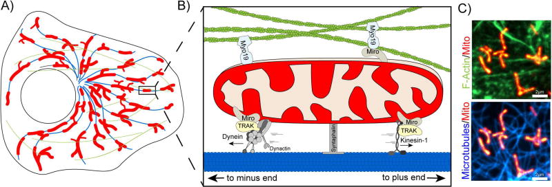
A. Schematic of mitochondria (red), microtubules (blue), and f-actin (green) distribution in an undifferentiated cell. B. Mitochondria associate with microtubules (blue, bottom) and with actin (green, top) via motor/adaptor complexes. Dynein/dynactin associate with mitochondria via TRAK and Miro to drive retrograde mitochondrial motility. In contrast, Kinesin-1 coordinates anterograde motility towards the cell periphery. Myo19 can associate with the mitochondria outer membrane either directly or through Miro. Syntaphilin anchors mitochondria to microtubules. C. Spinning disk confocal image of a HeLa cell expressing a mitochondria matrix marker (Mito-DsRed2) as w3ell as markers for filamentous actin (top, LifeAct-GFP) and microtubules (bottom, SiR-Tubulin).
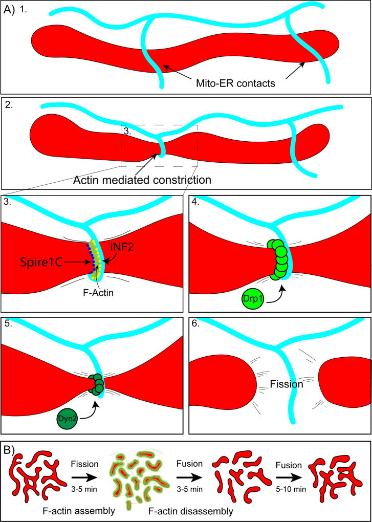
A) INF2/Spire1C mediated mitochondrial fission. 1. Mitochondria make frequent contacts with endoplasmic reticulum tubules (Mito-ER contacts). 2. A subset of Mito-ER contact sites mark prospective sites of mitochondrial fission. At these sites, ER tubules wrap around mitochondria generating a constriction event. 3. ER constriction around mitochondria is driven by actin polymerization by INF2 on the ER and Spire1C on mitochondria. 4. Once mitochondria undergo constriction, Drp1 forms rings at the constriction site which pinch down the organelle, decreasing the cross sectional diameter. 5. Next, Dyn2 assembles and completes the process of mitochondrial fission (6). B. Arp2/3 mediated mitochondrial fission. Actin transiently assembles on locally hyperfused regions of the mitochondrial network. Actin assembly promotes rapid fission over 3–5 minutes. Mitochondria then fuse back together and the process repeats.
References
-
- Schwarz TL. Mitochondrial trafficking in neurons. Mitochondria. 2014:163–178.
Grants and funding
LinkOut - more resources
Full Text Sources
Other Literature Sources