. 2019 Feb;12(2):200-211.
doi: 10.1002/aur.2051. Epub 2018 Dec 17.

PAC1R Genotype to Phenotype Correlations in Autism Spectrum Disorder

Affiliations

PAC1R Genotype to Phenotype Correlations in Autism Spectrum Disorder

Meredith Goodrich et al. Autism Res. 2019 Feb.

Abstract

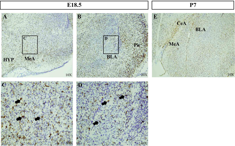
Amygdala dysfunction has been implicated in numerous neurodevelopmental disorders, including autism spectrum disorder (ASD). Previous studies in mice and humans, respectively, have linked Pac1r/PAC1R function to social behavior and PTSD-susceptibility. Based on this connection to social and emotional processing and the central role played by the amygdala in ASD, we examined a putative role for PAC1R in social deficits in ASD and determined the pattern of gene expression in the developing mouse and human amygdala. We reveal that Pac1r/PAC1R is expressed in both mouse and human amygdala from mid-neurogenesis through early postnatal stages, critical time points when altered brain trajectories are hypothesized to unfold in ASD. We further find that parents of autistic children carrying a previously identified PTSD-risk genotype (CC) report greater reciprocal social deficits compared to those carrying the non-risk GC genotype. Additionally, by exploring resting-state functional connectivity differences in a subsample of the larger behavioral sample, we find higher functional connectivity between the amygdala and right middle temporal gyrus in individuals with the CC risk genotype. Thus, using multimodal approaches, our data reveal that the amygdala-expressed PAC1R gene may be linked to severity of ASD social phenotype and possible alterations in brain connectivity, therefore potentially acting as a modifier of amygdala-related phenotypes. Autism Res 2019, 12: 200-211 © 2018 International Society for Autism Research, Wiley Periodicals, Inc. LAY SUMMARY: In this multimodal study across mouse and human, we examined expression patterns of Pac1r/PAC1R, a gene implicated in social behavior, and further explored whether a previously identified human PTSD-linked mutation in PAC1R can predict brain connectivity and social deficits in ASD. We find that PAC1R is highly expressed in the both the mouse and human amygdala. Furthermore, our human data suggest that PAC1R genotype is linked to severity of social deficits and functional amygdala connectivity in ASD.

Keywords: PAC1R; amygdala; autism; genetic modifier; neuroimaging.


Figures

Figure 1
Figure 1
ISH analyses at E13.5 shows Pac1r mRNA expression in known amygdala progenitor zones (A, boxed regions). Higher magnification image shows numerous Pac1r+ cells (brown) in the emerging amygdala (B) and the VP (C, arrows), a source of progenitors destined to give rise to excitatory output neurons in the BLA. Pac1r+ cells (arrow) are also observed in the POA (D), a source of progenitors destined to give rise to inhibitory output neurons in the MeA. Abbreviations: AMY, Amygdala; BLA, Basolateral Amygdala; CeA, Central Amygdala; CTX, Cortex; HIP, Hippocampus; MeA, Medial Amygdala; LGE, Lateral Ganglionic Eminence; MGE, Medial Ganglionic Eminence; POA, Pre-Optic Area; VP, Ventral Pallium.
Figure 2
Figure 2
ISH analyses at E18.5 shows high levels of Pac1r mRNA in developing amygdala nuclei (A, B). Higher magnification of the MeA (C) and BLA (D) show numerous Pac1r+ cells (arrows). (E) At post-natal day 7 many Pac1r+ cells are observed in multiple amygdala sub-nuclei. Abbreviations: BLA, basolateral amygdala; CeA, central nucleus of the amygdala; MeA, medial amygdala; HYP, hypothalamus; Pir, piriform cortex
Figure 3
Figure 3
Gene-level brain expression graphs were generated with data from the BrainSpan project (Kang et al., 2011) collected from post-mortem human brain samples. The x-axis shows periods of development with the dark vertical line signifying birth (developmental time points are defined in Table 1). Along the y-axis are values of median expression (Log2-Transformed Signal Intensity) for which values ≥ 6 signify high levels of expression in at least one post-mortem brain sample. A. Expression of PAC1R is observed in multiple brain regions starting at stage 1 of fetal development. B. Differential expression of PAC1R is observed between males and females in multiple brain regions with median values above 0.0 denoting higher expression in males and values below 0.0 denoting higher expression in females. C. Differential expression of PAC1R between males and females only in the amygdala. Abbreviations: AMY, amygdala; CBC, cerebellar cortex; HIP, hippocampus; MD, mediodorsal nucleus of the thalamus; NCX, neocortex; STR, striatum.
Figure 4
Figure 4
PAC1R is at a hub of different signaling cascades involved in stress, metabolism, and inflammatory responses, among others. Plot and legend adapted from Genomatix (v.s. 3.9).
Figure 5
Figure 5
ADI behavioral scores by PAC1R SNP allele status. A significant correlation exists between Reciprocal Social scores and allele status. Bars show standard error, n = 129, p < 0.05.
Figure 6
Figure 6
F test reveals one cluster in right middle temporal gyrus where functional connectivity to the bilateral amygdala was significantly higher in the CC group compared to the GC group. Bars show standard error, n = 20, p < 0.005.

References

    1. American Psychiatric Association. (2013). The diagnostic and statistical manual of mental disorders, 5th edition (DSM-5) Washington, DC: American Psychiatric Association.
    1. Barak B, & Feng G (2016). Neurobiology of social behavior abnormalities in autism and Williams syndrome. Nat Neurosci, 19(6). 647–655. doi: 10.1038/nn.4276. - DOI - PMC - PubMed
    1. Baron-Cohen S, Ring HA, Bullmore ET, Wheelwright S, Ashwin C, & Williams SCR (2000). The amygdala theory of autism. Neuroscience & Biobehavioral Reviews, 24(3), 355–364. 10.1016/S0149-7634(00)00011-7 - DOI - PubMed
    1. Bindea G, Mlecnik B, Hackl H, Charoentong P, Tosolini M, Kirilovsky A, … Galon J (2009). ClueGO: a Cytoscape plug-in to decipher functionally grouped gene ontology and pathway annotation networks. Bioinformatics, 25(8), 1091–1093. 10.1093/bioinformatics/btp101 - DOI - PMC - PubMed
    1. Bozzi Y, Provenzano G, & Casarosa S (2018). Neurobiological bases of autism-epilepsy comorbidity: a focus on excitation/inhibition imbalance. Eur J Neurosci, 47(6), 534–548. doi: 10.1111/ejn.13595. - DOI - PubMed

Publication types

Substances