Review
. 2019 Jan;8(2):e1801236.
doi: 10.1002/adhm.201801236. Epub 2018 Dec 17.

Tissue Engineering for the Temporomandibular Joint

Affiliations
Review

Tissue Engineering for the Temporomandibular Joint

Timothy M Acri et al. Adv Healthc Mater. 2019 Jan.

Abstract

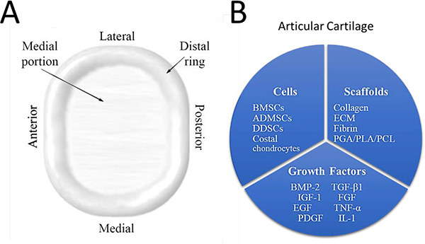
Tissue engineering potentially offers new treatments for disorders of the temporomandibular joint which frequently afflict patients. Damage or disease in this area adversely affects masticatory function and speaking, reducing patients' quality of life. Effective treatment options for patients suffering from severe temporomandibular joint disorders are in high demand because surgical options are restricted to removal of damaged tissue or complete replacement of the joint with prosthetics. Tissue engineering approaches for the temporomandibular joint are a promising alternative to the limited clinical treatment options. However, tissue engineering is still a developing field and only in its formative years for the temporomandibular joint. This review outlines the anatomical and physiological characteristics of the temporomandibular joint, clinical management of temporomandibular joint disorder, and current perspectives in the tissue engineering approach for the temporomandibular joint disorder. The tissue engineering perspectives have been categorized according to the primary structures of the temporomandibular joint: the disc, the mandibular condyle, and the glenoid fossa. In each section, contemporary approaches in cellularization, growth factor selection, and scaffold fabrication strategies are reviewed in detail along with their achievements and challenges.

Keywords: animal studies; cells; growth factors; scaffolds; temporomandibular joint; tissue engineering.


Figures

Figure 1.
Figure 1.
Anatomic visualization of the TMJ.
Figure 2.
Figure 2.
Anatomy and tissue engineering strategies for the articular disc. Anatomy of the articular disc (A), and attempted tissue engineering strategies specific for the disc (B).
Figure 3
Figure 3
Histology and qPCR data for DPSCs in chondrogenic media. Imaging for cartilage deposition in the ECM was performed using hematoxylin and eosin stain, alcian blue, and immunohistochemistry for aggrecan. DPSCs cultured in chondrogenic media for 14 days were assess using qPCR for chondrogenic markers Sex determining region Y-box 9 [Sox9], Collagen I [COL I], Collagen II [COL X], Aggrecan [ACAN], Cartilage Oligomeric Matrix Protein [COMP] and compared to the baseline of day 7 gene expression. Scale bars are 50 μm; error bars represent SD. Modified from “Fibro/chondrogenic differentiation of dental stem cells into chitosan/alginate scaffolds towards temporomandibular joint disc regeneration” by Bousnaki et al. with permission from Springer Nature, 2018[73].
Figure 4.
Figure 4.
Self-assembled cartilage constructs implanted in a minipig TMJ disc perforation model assessed after eight weeks. Histology (A), defect perimeter closure (B), mechanical testing (C), and osteoarthritis [OA] score (D) all indicate the tissue engineer [TE] implant group improved wound healing. Scale bar is 2 mm; error bars represent SD. Reproduced from “Tissue engineering toward temporomandibular joint disc regeneration” by Vapniarsky et al. with permission from AAAS, 2018[83].
Figure 5.
Figure 5.
TMJ-SDSCs seeded on fibrin/chitosan scaffolds implanted in a murine model for four weeks. Immunohistochemical staining for collagen I (Col I) and collagen II (Col II) was performed demonstrating more collagen was deposited in the fibrin-coated scaffold (A). Additionally, cell viability testing (B) and qPCR for collage I and collagen II (C) were performed. Error bars represent SD and asterisks indicate P < 0.05. Reproduced from “The Pilot Study of Fibrin with Temporomandibular Joint Derived Synovial Stem Cells in Repairing TMJ Disc Perforation” by Wu et al. under the CC BY 2.0, 2014[65].
Figure 6.
Figure 6.
3D printed PCL scaffold embedded with protein-loaded microspheres for TMJ disc regeneration. Laser scan and 3D print of the TMJ disc (A, B). The tensile modulus of the 3D printed scaffolds compared to the native TMJ disc (C). Fluorescently labeled particles embedded in a scaffold to demonstrate spatiocontrol (D). The release of the growth factors from the scaffold (E). Reproduced from “Engineering human TMJ discs with protein-releasing 3D-printed scaffolds” by Legemate et al. with permission from SAGE Publications, 2016[2].
Figure 7.
Figure 7.
Anatomy and tissue engineering strategies for the mandibular condyle. Anatomy of the mandibular condyle (A), and attempted tissue engineering strategies specific for each tissue type present in the mandibular condyle (B).
Figure 8.
Figure 8.
Single FCSC expanded, seeded on a collagen sponge, and implanted in the dorsum of nude mice (A). After three weeks H&E staining and immunohistochemistry staining for aggrecan (ACAN) revealed cartilage formation (B). Six weeks post-implantation, H&E staining and immunohistochemistry staining for osteocalcin (OCN) revealed bone formation (C) Scale bars are 50 μm. Reproduced from “Exploiting endogenous fibrocartilage stem cells to regenerate cartilage and repair joint injury” by Embree et al. under CC BY 4.0, 2016[141].
Figure 9.
Figure 9.
Injection of VEGF adenovirus gene therapy into the TMJ. Protein expression for alkaline phosphatase (A) and osteocalcin (B) were upregulated in the treated group at 28 days Asterisks represent *P < 0.05 and **P < 001 Reproduced from “Recombinant AAV-mediated VEGF gene therapy induces mandibular condylar growth” by Rabie et al. with permission from Springer Nature and Copyright Clearance Center, 2007[1].
Figure 10.
Figure 10.
Biphasic scaffold for osteochondral integration. The PLG/PLA cartilage scaffold (A) was sutured to the superior surface of the 3D printed HA/PCL scaffold (B) to form the biphasic scaffold (C). After 12 weeks of implantation, gross morphology (D), H&E staining (E), and safranin staining (F) were performed. The cartilaginous area [C] and the cartilage-bone interface [i] are indicated in panel E&F. Reproduced from “Regeneration of subcutaneous tissue-engineered mandibular condyle in nude mice” by Wang et al. with permission from Elsevier and Copyright Clearance Center, 2017[178].
Figure 11.
Figure 11.
Anatomy and potential tissue engineering strategies for the glenoid fossa and articular eminence. Anatomy of the glenoid fossa and articular eminence (A), and attempted tissue engineering strategies specific for each tissue type present in the glenoid fossa and articular eminence (B).
Figure 12.
Figure 12.
3D printed PCL scaffolds seeded with BMSCs for knee meniscus repair in a rabbit model. The scaffolds were implanted for 24 weeks, and gross morphology (upper row; scale bar is 10 mm) and toluidine blue staining (low row; scale bar is 100 μm) were performed (A). The native tissue and the implanted scaffolds are indicated as [N] and [S] respectively. Immunohistochemical staining was performed and quantified demonstrating collagen I (Col I) (B) was upregulated at 24 weeks whereas collagen II (Col II) (C) was significantly upregulated at 12 and 24 weeks in the cell-seeded scaffolds. Error bars represent SD; asterisks represent *** P < 0.001. Reproduced from “3D-printed poly(epsilon-caprolactone) scaffold augmented with mesenchymal stem cells for total meniscal substitution: a 12- and 24-week animal study in a rabbit model” by Zhang et al. with permission from SAGE Publications, 2017[228].

References

    1. Rabie ABM, Dai J, and Xu R, Recombinant AAV-mediated VEGF gene therapy induces mandibular condylar growth. Gene Therapy, 2007. 14: p. 972. - PubMed
    1. Legemate K, et al., Engineering Human TMJ Discs with Protein-Releasing 3D-Printed Scaffolds. Journal of Dental Research, 2016. 95(7): p. 800–807. - PubMed
    1. Sharma S, et al., Etiological factors of temporomandibular joint disorders. Natl J Maxillofac Surg, 2011. 2(2): p. 116–9. - PMC - PubMed
    1. Dorland WAN, et al., Dorland’s illustrated medical dictionary. 1957, Philadelphia; London: W. B. Saunders Co.
    1. Helland MM, Anatomy and function of the temporomandibular joint. J Orthop Sports Phys Ther, 1980. 1(3): p. 145–52. - PubMed

Publication types

MeSH terms

Substances