

Regulation of Protein Activity and Cellular Functions Mediated by Molecularly Evolved Nucleic Acids
- PMID: 30556364
- PMCID: PMC6442720
- DOI: 10.1002/anie.201809010
Regulation of Protein Activity and Cellular Functions Mediated by Molecularly Evolved Nucleic Acids
Abstract
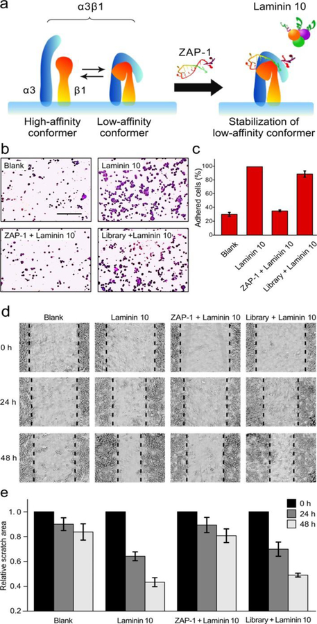
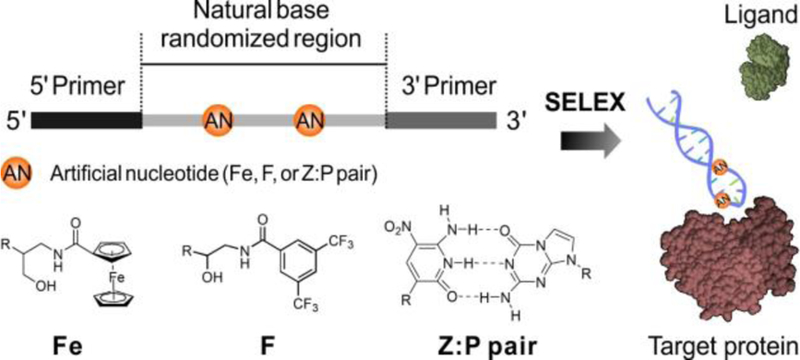
Regulation of protein activity is essential for revealing the molecular mechanisms of biological processes. DNA and RNA achieve many uniquely efficient functions, such as genetic expression and regulation. The chemical capability to synthesize artificial nucleotides can expand the chemical space of nucleic acid libraries and further increase the functional diversity of nucleic acids. Herein, a versatile method has been developed for modular expansion of the chemical space of nucleic acid libraries, thus enabling the generation of aptamers able to regulate protein activity. Specifically, an aptamer that targets integrin alpha3 was identified and this aptamer can inhibit cell adhesion and migration. Overall, this chemical-design-assisted in vitro selection approach enables the generation of functional nucleic acids for elucidating the molecular basis of biological activities and uncovering a novel basis for the rational design of new protein-inhibitor pharmaceuticals.
Keywords: aptamers; cell adhesion; inhibitors; membrane proteins; molecular evolution.
© 2019 Wiley-VCH Verlag GmbH & Co. KGaA, Weinheim.
Conflict of interest statement
Conflict of interest
The authors declare no conflict of interest.
Figures

References
-
- a) Ellington AD, Szostak JW, Nature 1992, 355, 850. - PubMed
- b) Bock LC, Griffin LC, Latham JA, Vermaas EH, Toole JJ, Nature 1992, 355, 564. - PubMed
- c) Kim YM, Phillips JA, Liu HP, Kang HZ, Tan WH, Proc. Natl. Acad. Sci. USA 2009, 106, 6489. - PMC - PubMed
- d) Wang J, Wei YR, Hu XX, Fang YY, Li XY, Liu J, Wang SF, Yuan Q, J. Am. Chem. Soc 2015, 137, 10576. - PubMed
-
- a) Lao YH, Phua KKL, Leong KW, Acs Nano 2015, 9, 2235. - PubMed
- b) Simona C, Laura C, Cent. Nerv. Syst. Agents Med. Chem 2015, 15, 126. - PubMed
- c) Zhou JH, Rossi J, Nat. Rev. Drug Discov 2017, 16, 181. - PMC - PubMed
- d) Zhuo ZJ, Yu YY, Wang ML, Li J, Zhang ZK, Liu J, Wu XH, Lu AP, Zhang G, Zhang BT, Int. J. Mol. Sci 2017, 18, 2430 - PMC - PubMed
- e) Röthlisberger P, Gasse C, Hollenstein M, Int. J. Mol. Sci 2017, 18, 2142. - PMC - PubMed
Publication types
MeSH terms
Substances
Grants and funding
- 21327009/National Natural Science Foundation of China/International
- 21675120/National Natural Science Foundation of China/International
- 531107050973/National Key R&D Program of China/International
- 21521063/National Natural Science Foundation of China/International
- R35 GM127130/GM/NIGMS NIH HHS/United States
LinkOut - more resources
Full Text Sources
