. 2018 Dec 17;14(12):e1007828.
doi: 10.1371/journal.pgen.1007828. eCollection 2018 Dec.

The Integrator complex regulates differential snRNA processing and fate of adult stem cells in the highly regenerative planarian Schmidtea mediterranea

Affiliations

The Integrator complex regulates differential snRNA processing and fate of adult stem cells in the highly regenerative planarian Schmidtea mediterranea

David Schmidt et al. PLoS Genet. .

Abstract

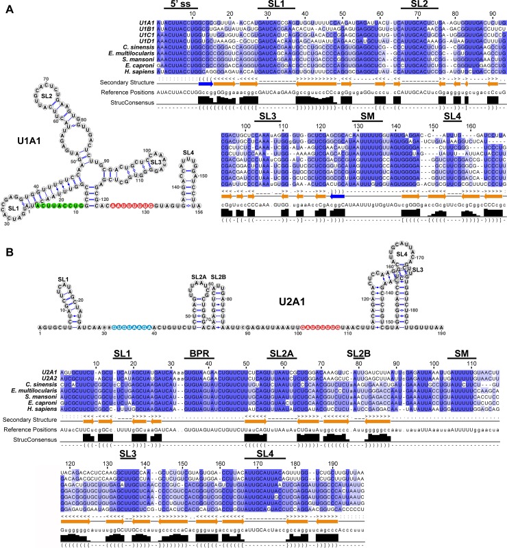
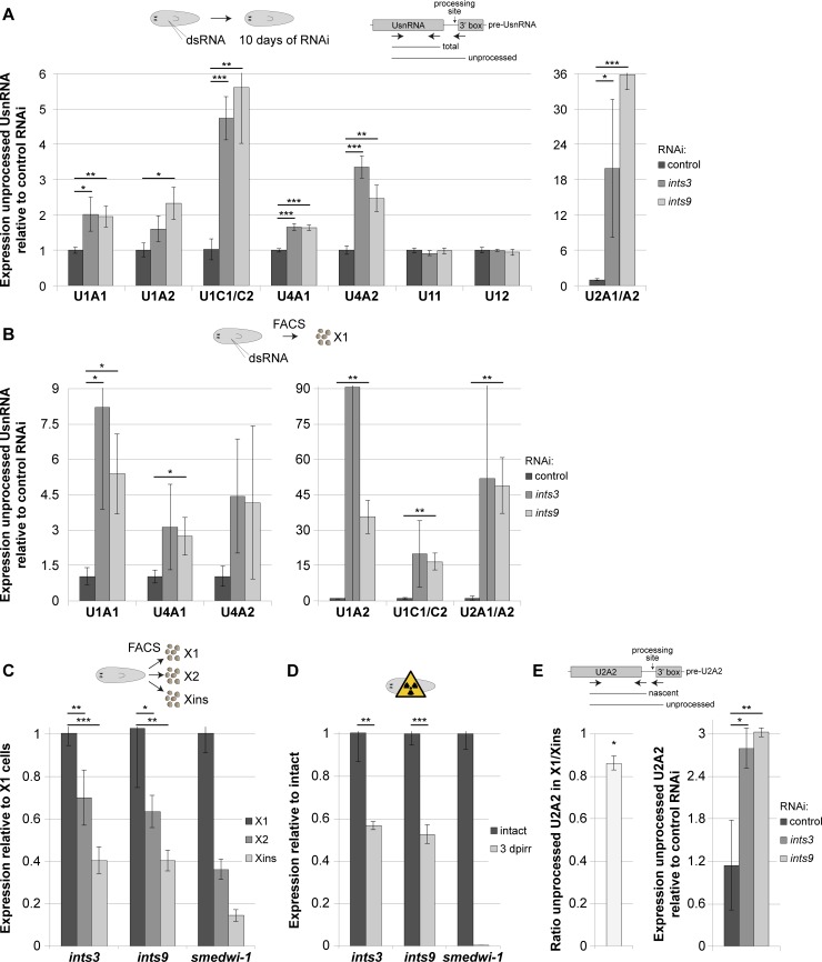
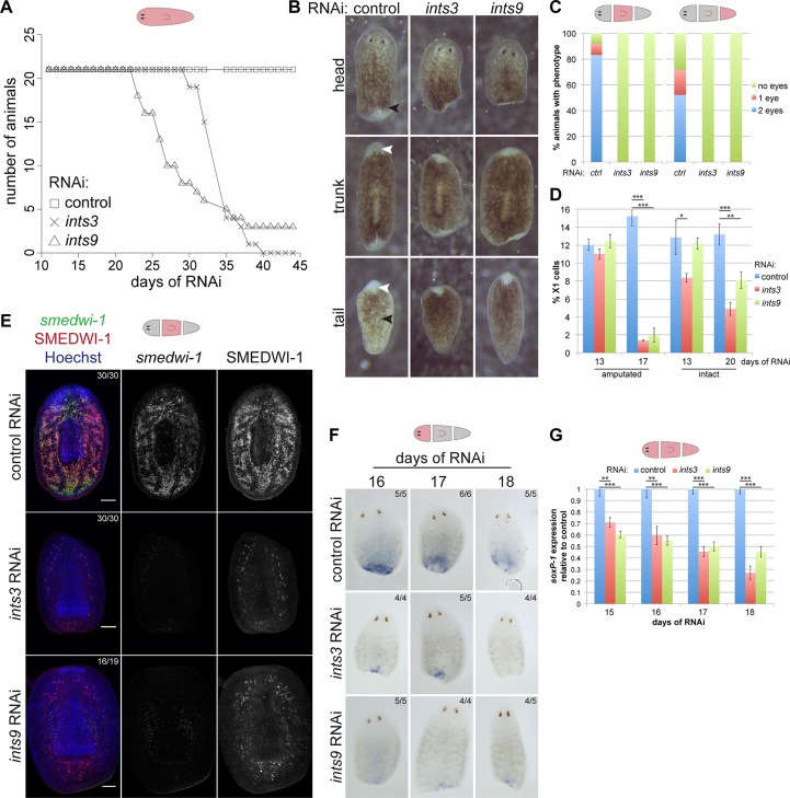
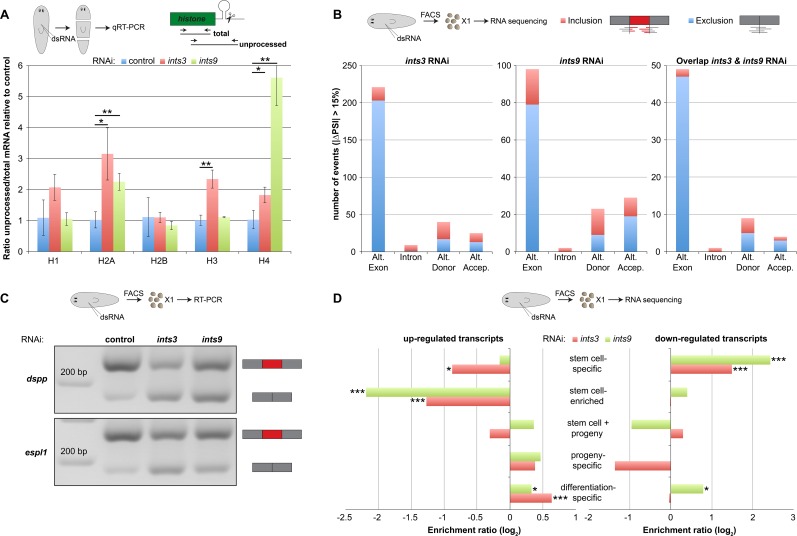
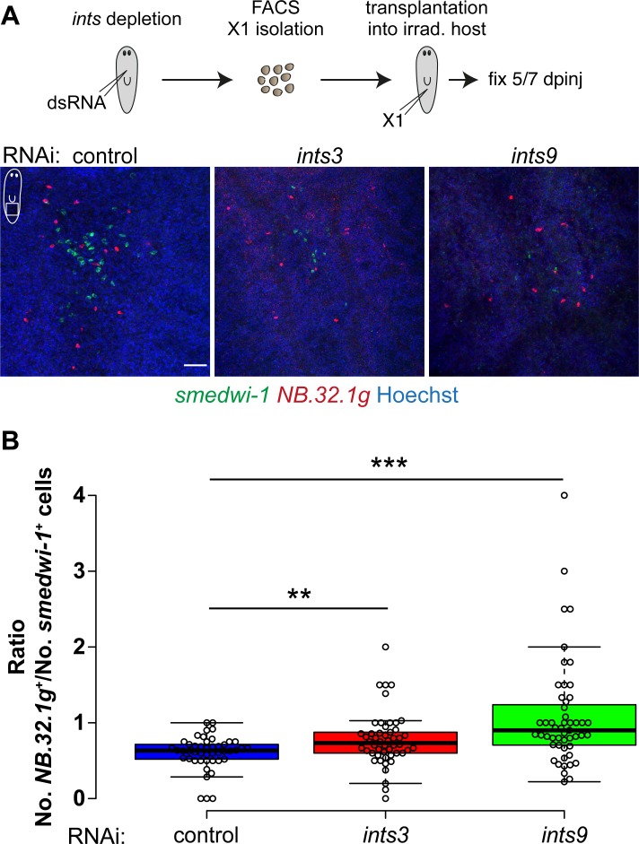
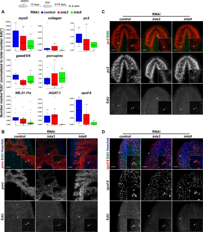
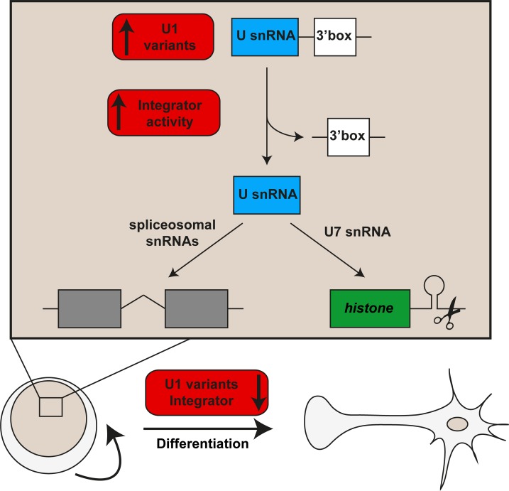
In multicellular organisms, cell type diversity and fate depend on specific sets of transcript isoforms generated by post-transcriptional RNA processing. Here, we used Schmidtea mediterranea, a flatworm with extraordinary regenerative abilities and a large pool of adult stem cells, as an in vivo model to study the role of Uridyl-rich small nuclear RNAs (UsnRNAs), which participate in multiple RNA processing reactions including splicing, in stem cell regulation. We characterized the planarian UsnRNA repertoire, identified stem cell-enriched variants and obtained strong evidence for an increased rate of UsnRNA 3'-processing in stem cells compared to their differentiated counterparts. Consistently, components of the Integrator complex showed stem cell-enriched expression and their depletion by RNAi disrupted UsnRNA processing resulting in global changes of splicing patterns and reduced processing of histone mRNAs. Interestingly, loss of Integrator complex function disrupted both stem cell maintenance and regeneration of tissues. Our data show that the function of the Integrator complex in UsnRNA 3'-processing is conserved in planarians and essential for maintaining their stem cell pool. We propose that cell type-specific modulation of UsnRNA composition and maturation contributes to in vivo cell fate choices, such as stem cell self-renewal in planarians.


Conflict of interest statement

The authors have declared that no competing interests exist.

Figures

Fig 1
Fig 1. Features of Schmidtea mediterranea major spliceosomal UsnRNAs.
Representative predicted secondary structures of (A) U1 and (B) U2 snRNAs and sequence alignments to their homologs of other Platyhelminthes (Clonorchis sinensis, Echinococcus multilocularis, Schistosoma mansoni and Echinostoma caproni) and Homo sapiens. Primary sequence similarity is indicated in violet and secondary structure conservation (StrucConsensus) is shown below the alignments. Evolutionary conserved structural features and functional sequence elements are indicated: 5’ ss = 5’ splice site recognition motif (green); SL = stem loop; SM = SM protein binding site (red); BPR = branch-point recognition sequence (blue).
Fig 2
Fig 2. Neoblast-enriched expression of variant U1 snRNAs.
(A, B) UsnRNA variant expression, quantified by qRT-PCR, in animals depleted of neoblasts by h2b RNAi (blue) or lethal irradiation (green) and in cell populations isolated by FACS (red). Shown are the variants with significantly changed expression relative to the respective control. Results for other variants are shown in S3 Fig. Expression levels are the averages of three biological replicates, normalized to those of gapdh (error bars represent standard deviation; two-sided t-test, * p<0.05, ** p<0.01, *** p<0.001). (C) Predicted secondary structures and (D) sequence alignment of S. mediterranea U1 snRNA variants. Evolutionary conserved structural features and functional sequence elements are indicated: 5’ ss = 5’ splice site recognition motif (green); SL = stem loop; SM = SM protein binding site (red). Primary sequence similarity is indicated in violet and secondary structure conservation (StrucConsensus) is shown below the alignments.
Fig 3
Fig 3. Expression of integrator genes is increased in neoblasts and is essential for UsnRNA processing.
qRT-PCR quantification of transcript levels. (A, B) Depletion of Integrator complex subunits results in a significant accumulation of 3’-unprocessed UsnRNAs in both intact animals and in the neoblast-enriched X1 FACS fraction. (C, D) ints3 and ints9 mRNA levels were significantly higher in the X1 fraction compared to both the X2 and the Xins fraction, and were strongly down-regulated in γ-irradiated animals depleted of neoblasts. (E) 3’-unprocessed U2A2 snRNA, relative to the total nascent U2A2 level, was less abundant in RNA of X1 cells compared to that of Xins cells (left chart; average of the X1/Xins ratios of 3 biological replicates; p = 0.02, two-sided heteroscedastic t-test) and accumulated substantially in X1 cells upon ints RNAi (right chart). Expression levels are the averages of three biological replicates normalized to those of the total pool of the respective UsnRNA variant in A+B, to those of gapdh in C+D and to those of total nascent U2A2 in E (error bars represent standard deviation; two-sided t-test, * p<0.05, ** p<0.01, *** p<0.001).
Fig 4
Fig 4. ints depletion disrupts neoblast maintenance as well as tissue homeostasis and regeneration.
(A) Survival curves of homeostatic ints RNAi animals showing median survival times of 32 and 26 days after the first RNAi treatment (days of RNAi) for ints3 and ints9 RNAi, respectively. Corresponding live images are in S4A Fig. (B) Representative live images of ints RNAi fragments 7 days post amputation (7 dpa; 20 days RNAi) displaying significantly reduced blastema size, lack of eye regeneration (white arrowheads) and defective remodeling of existing tissues to form a pharynx (black arrowheads). (C) Quantification of eye regeneration in trunk and tail fragments corresponding to B (n>32; 2 independent experiments). (D) Cytometry analysis of dissociated cells derived from ints and control RNAi animals revealing a progressive loss of the X1 populations in both amputated and intact ints RNAi animals. Shown is the average proportion of X1 cells in all gated cells of 3 biological replicates. (E) FISH and immunostaining of smedwi-1 mRNA and SMEDWI-1 protein, respectively, in trunk fragments at 7 dpa (20 days of RNAi) confirming the ints RNAi-induced loss of neoblasts and their immediate progeny. The proportion of trunk fragments with the depicted phenotype are indicated (2 independent experiments; Scale bar = 100 μm). (F+G) WISH staining- and qRT-PCR time-course (2–5 dpa) showing that the expression of soxP-1, a marker of sigma neoblasts, is strongly reduced in ints RNAi fragments. The expression levels are the averages of three biological replicates normalized to those of gapdh. (two-sided t-test, * p<0.05, ** p<0.01, *** p<0.001). Error bars represent standard deviation.
Fig 5
Fig 5. ints RNAi interferes with histone mRNA processing and disrupts the splicing- and gene expression profiles of neoblasts.
(A) qRT-PCR quantification showing that the 3’-unprocessed mRNAs of H2A, H3 and H4 accumulate in regenerating ints RNAi animals (3 dpa; 16 days of RNAi). Expression levels are the averages of three biological replicates normalized to those of the total pool of the respective histone (Error bars represent standard deviation; two-sided t-test, * p<0.05, ** p<0.01). (B) Overview of the splicing events significantly changed in neoblasts depleted of ints3, ints9 or in both conditions showing that the exclusion of alternative (Alt.) exons (exon skipping) is the primary splicing defect induced by ints RNAi. PSI = Percent Spliced-In. For details see S4 Table. (C) Two examples of ints RNAi-induced exon skipping events in neoblasts affecting transcripts encoding putative homologs of dspp and espl1, analyzed by RT-PCR. Results for all other RT-PCR validations are shown in S7A Fig. (D) Comparison of the gene expression changes induced by ints RNAi in X1 cells to the transcripts differentially expressed between the X1, X2 and Xin FACS fractions [41]. Transcripts annotated as stem cell-specific were statistically significantly over-represented among the transcripts down-regulated in ints RNAi X1 cells (threshold for differentially expressed genes in ints RNAi neoblasts: padj<0.05; |log2 fold-change| > 1; Statistical test for over-/under-representation: hypergeometric test, * p<0.05, *** p<0.001).
Fig 6
Fig 6. ints depletion disrupts neoblast self-renewal.
(A) Neoblast transplantation assay. FACS-isolated X1 cells of RNAi animals were transplanted into neoblast-depleted, wild-type hosts. The cell type composition of colonies derived from transplanted neoblasts was analyzed by FISH staining of the neoblast- and the early progeny marker smedwi-1 and NB.32.1g, respectively. ints-depleted neoblasts produced smaller colonies containing an increased proportion of early progeny. Scale bar = 50 μm. dpinj = days post neoblast injection. (B) Quantification of colony composition showing a significant increase in the proportion of early progeny (NB.32.1g+) cells generated from ints-depleted neoblasts. Data of three independent experiments. Circles represent individual colonies. Center lines show the medians; box limits indicate the 25th and 75th percentiles as determined by R software; whiskers extend 1.5 times the interquartile range from the 25th and 75th percentiles, outliers are represented by dots. n = 46, 50, 51 colonies. Two-sided t-test: ** p<0.01, *** p<0.001.
Fig 7
Fig 7. ints RNAi reduces differentiated cell output.
EdU pulse-chase labeling combined with FISH staining of differentiation markers. 10 days following the first dsRNA injection, animals were pulsed with EdU, fixed 3, 7 or 9 days later and cell type markers were visualized by FISH. (A) Quantification of EdU-positive cells expressing the indicated marker, normalized to the total number of EdU-positive cells. Chase periods were 3 days for NB.21.11.e, myoD and gata4/5/6, 7 days for AGAT-1 and 9 days for porcupine, pc2, zpuf-6 and collagen. Circles represent measurements of individual animals. Center lines show the medians; box limits indicate the 25th and 75th percentiles as determined by R software; whiskers extend 1.5 times the interquartile range from the 25th and 75th percentiles, outliers are represented by dots. (B, C, D) Representative images of EdU-labelled cells expressing the intestinal (porcupine), neuronal (pc2) or epidermal progeny (zpuf-6) marker. Overview images and insets are single confocal sections acquired with a 20x and a 63x objective, respectively. Arrowheads indicate marker+EdU+ cells. Scale bars: overview pictures = 50 μm, magnifications = 5 μm.
Fig 8
Fig 8. Model of the role of UsnRNAs in neoblast maintenance.
Neoblasts express higher levels of U1 snRNA variants and have higher UsnRNA 3’-processing activity, likely due to the high expression levels of Integrator complex components. Together with increased expression of spliceosomal proteins [14], this may result in a modified specificity and enhanced activity of the spliceosome in neoblasts compared to differentiated cell types. In addition, an increased activity of the Integrator complex may allow efficient histone mRNA 3’-processing through U7 snRNA maturation. Together, these features likely contribute to neoblast maintenance by generating a specific set of protein isoforms as well as by enabling the high turnover of mRNAs and proteins in these mitotically active cells.

Similar articles

Cited by

References

    1. Schöler HR, Dressler GR, Balling R, Rohdewohld H, Gruss P. Oct-4: a germline-specific transcription factor mapping to the mouse t-complex. EMBO J. 1990;9: 2185–2195. - PMC - PubMed
    1. Rosner MH, Vigano MA, Ozato K, Timmons PM, Poirier F, Rigby PW, et al. A POU-domain transcription factor in early stem cells and germ cells of the mammalian embryo. Nature. 1990;345: 686–692. 10.1038/345686a0 - DOI - PubMed
    1. Takahashi K, Yamanaka S. Induction of pluripotent stem cells from mouse embryonic and adult fibroblast cultures by defined factors. Cell. 2006;126: 663–676. 10.1016/j.cell.2006.07.024 - DOI - PubMed
    1. Gabut M, Samavarchi-Tehrani P, Wang X, Slobodeniuc V, O’Hanlon D, Sung H-K, et al. An alternative splicing switch regulates embryonic stem cell pluripotency and reprogramming. Cell. 2011;147: 132–146. 10.1016/j.cell.2011.08.023 - DOI - PubMed
    1. Han H, Irimia M, Ross PJ, Sung H-K, Alipanahi B, David L, et al. MBNL proteins repress ES-cell-specific alternative splicing and reprogramming. Nature. 2013;498: 241–245. 10.1038/nature12270 - DOI - PMC - PubMed

Publication types

MeSH terms