

The predictive value of Cardiodynamicsgram in myocardial perfusion abnormalities
- PMID: 30557346
- PMCID: PMC6296510
- DOI: 10.1371/journal.pone.0208859
The predictive value of Cardiodynamicsgram in myocardial perfusion abnormalities
Abstract
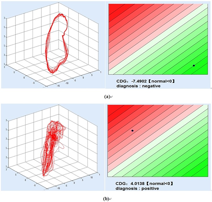
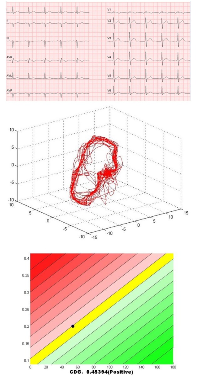
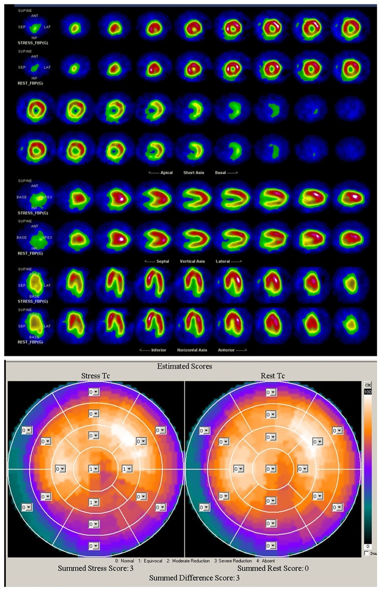
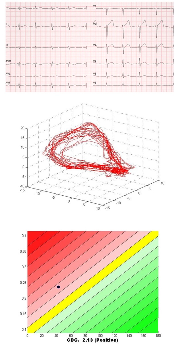
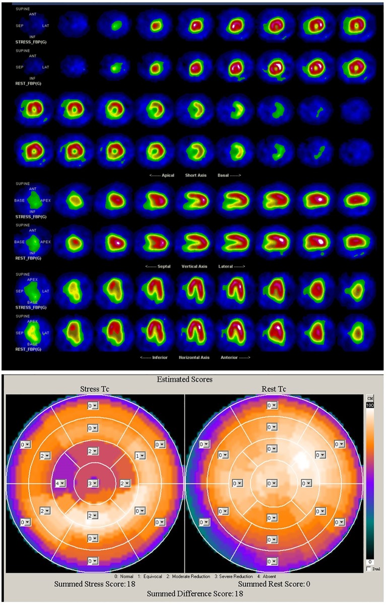
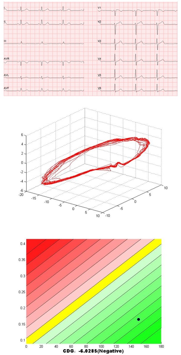
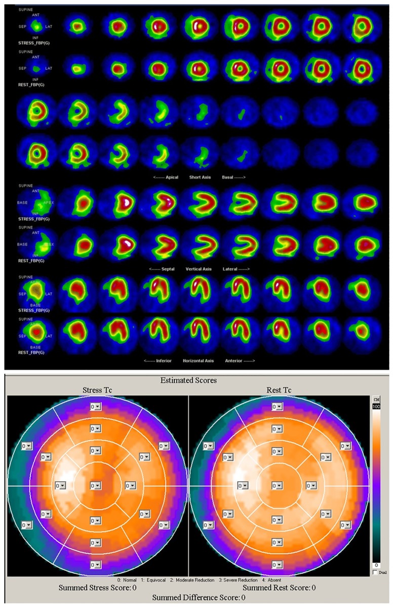
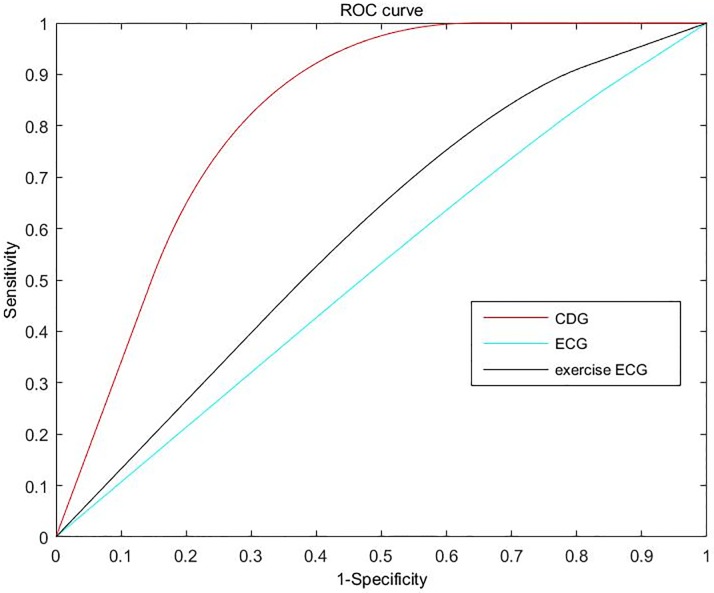
Myocardial perfusion abnormalities are the first sign of the ischemic cascade in the development of coronary artery disease (CAD). Thus, the early detection of myocardial perfusion abnormalities is significant for the prevention of CAD. Recently, a novel noninvasive method named Cardiodynamicsgram (CDG) has been proposed for early detection of CAD. This study aims to evaluate the predictive value of CDG in myocardial perfusion abnormalities for suspected ischemic heart disease. In the study, 86 suspected patients were enrolled. Standard 12-lead ECG and CDG were performed simultaneously before single-photon emission computed tomography (SPECT) myocardial perfusion imaging (MPI). Diagnostic accuracy of CDG for myocardial perfusion abnormalities detection is assessed using SPECT MPI as the reference standard. Of these 86 suspected patients, 37 patients were positive in CDG, 49 patients were negative in CDG. Diagnostic accuracy of CDG at presentation for myocardial perfusion abnormalities was 84.9%, sensitivity 84.0%, and specificity 89.4%. Furthermore, of the 10 patients whose SPECT MPI results are reverse redistribution, 9 patients were positive in CDG. Underlying causes of false positive CDG findings included the factors that can change the stability of cardiac electrical conduction and measurement noise. Myocardial remodeling in patients with old myocardial infarction might be the major cause of false negative findings. Results show a good consistency between the CDG and SPECT MPI in evaluating myocardial perfusion abnormalities. It suggests that CDG might be used as a cost-effective tool for assessing the myocardial perfusion abnormalities in the clinic.
Conflict of interest statement
The authors have declared that no competing interests exist.
Figures
Similar articles
-
Cardiodynamicsgram as a New Diagnostic Tool in Coronary Artery Disease Patients With Nondiagnostic Electrocardiograms.Am J Cardiol. 2017 Mar 1;119(5):698-704. doi: 10.1016/j.amjcard.2016.11.028. Epub 2016 Dec 2. Am J Cardiol. 2017. PMID: 28017302
-
Ischemic burden assessment of myocardial perfusion CT, compared with SPECT using semi-quantitative and quantitative approaches.Int J Cardiol. 2019 Mar 1;278:287-294. doi: 10.1016/j.ijcard.2018.12.046. Epub 2018 Dec 17. Int J Cardiol. 2019. PMID: 30587418 Clinical Trial.
-
Additional diagnostic value of integrated analysis of cardiac CTA and SPECT MPI using the SMARTVis system in patients with suspected coronary artery disease.J Nucl Med. 2014 Jan;55(1):50-7. doi: 10.2967/jnumed.113.119842. Epub 2013 Dec 12. J Nucl Med. 2014. PMID: 24337600
-
Serial myocardial perfusion imaging: defining a significant change and targeting management decisions.JACC Cardiovasc Imaging. 2014 Jan;7(1):79-96. doi: 10.1016/j.jcmg.2013.05.022. JACC Cardiovasc Imaging. 2014. PMID: 24433711 Review.
-
Physiological assessment of myocardial perfusion using nuclear cardiology would enhance coronary artery disease patient care: which imaging modality is best for evaluation of myocardial ischemia? (SPECT-side).Circ J. 2011;75(3):713-22; discussion 731. doi: 10.1253/circj.cj-10-1290. Epub 2011 Feb 3. Circ J. 2011. PMID: 21301136 Review.
Cited by
-
Early and Accurate Detection of Radiation-induced Heart Damage by Cardiodynamicsgram.J Cardiovasc Transl Res. 2024 Apr;17(2):242-251. doi: 10.1007/s12265-023-10419-0. Epub 2023 Aug 7. J Cardiovasc Transl Res. 2024. PMID: 37548860
-
Reliable Detection of Myocardial Ischemia Using Machine Learning Based on Temporal-Spatial Characteristics of Electrocardiogram and Vectorcardiogram.Front Physiol. 2022 May 30;13:854191. doi: 10.3389/fphys.2022.854191. eCollection 2022. Front Physiol. 2022. PMID: 35707012 Free PMC article.
-
ECG-based cardiodynamicsgram can reflect anomalous functional information in coronary artery disease.Clin Cardiol. 2023 Jun;46(6):639-647. doi: 10.1002/clc.24019. Epub 2023 Apr 6. Clin Cardiol. 2023. PMID: 37025093 Free PMC article.
References
-
- Organization WH. Cardiovascular diseases (CVDs); 2017. Available from: http://www.who.int/mediacentre/factsheets/fs317/en/.
-
- Qayyum A, Kastrup J. Measuring myocardial perfusion: the role of PET, MRI and CT. Clinical radiology. 2015;70(6):576–584. 10.1016/j.crad.2014.12.017 - DOI - PubMed
-
- Tung R, Bauer B, Schelbert H, Lynch JP III, Auerbach M, Gupta P, et al. Incidence of abnormal positron emission tomography in patients with unexplained cardiomyopathy and ventricular arrhythmias: the potential role of occult inflammation in arrhythmogenesis. Heart Rhythm. 2015;12(12):2488–2498. 10.1016/j.hrthm.2015.08.014 - DOI - PMC - PubMed
-
- Barizon GC, Simoes MV, Schmidt A, Gadioli LP, Murta LO Junior. Relationship between microvascular changes, autonomic denervation, and myocardial fibrosis in Chagas cardiomyopathy: Evaluation by MRI and SPECT imaging. Journal of Nuclear Cardiology. 2018; p. 1–11. - PubMed
-
- Beller GA. Future growth and success of nuclear cardiology. Journal of Nuclear Cardiology. 2018;25(2):375–378. 10.1007/s12350-018-1211-1 - DOI - PubMed
Publication types
MeSH terms
LinkOut - more resources
Full Text Sources
Medical
Miscellaneous
