

The predictive value of Cardiodynamicsgram in myocardial perfusion abnormalities
- PMID: 30557346
- PMCID: PMC6296510
- DOI: 10.1371/journal.pone.0208859
The predictive value of Cardiodynamicsgram in myocardial perfusion abnormalities
Abstract
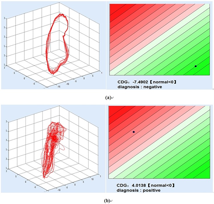
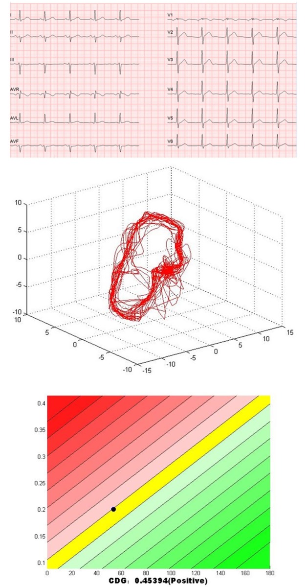
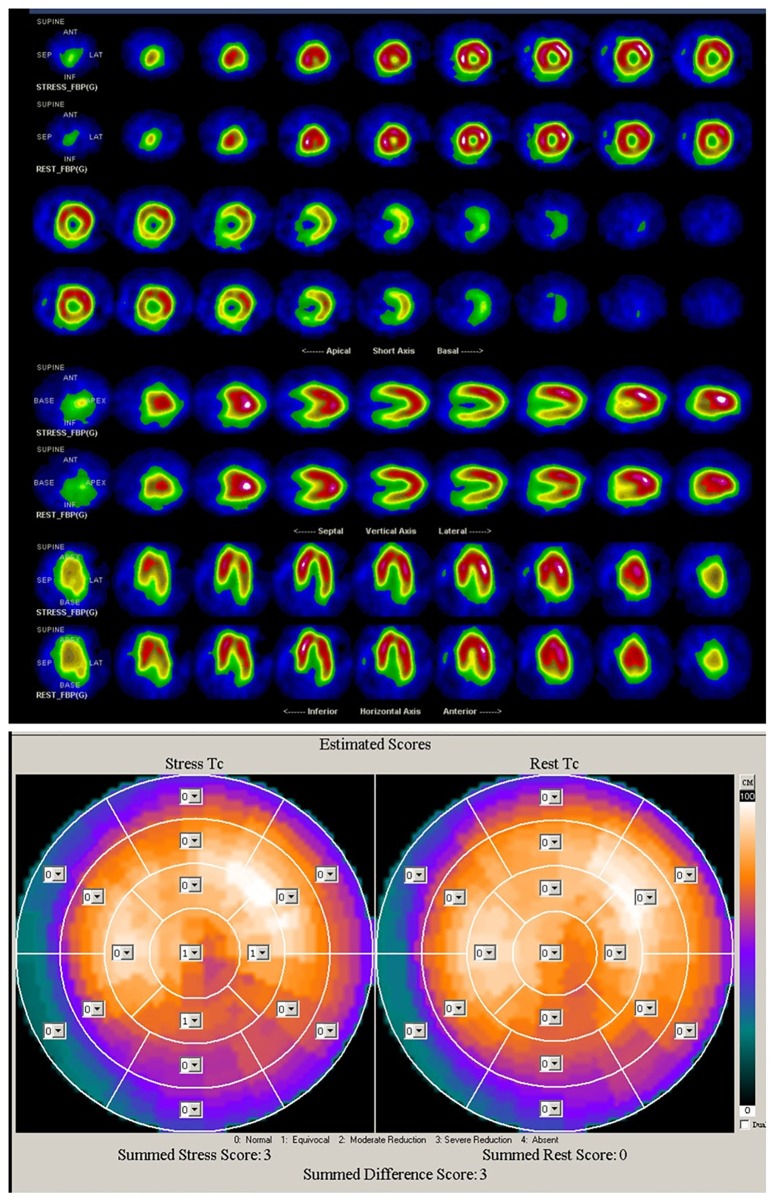
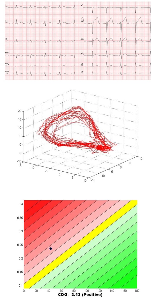
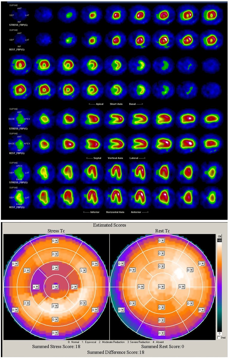
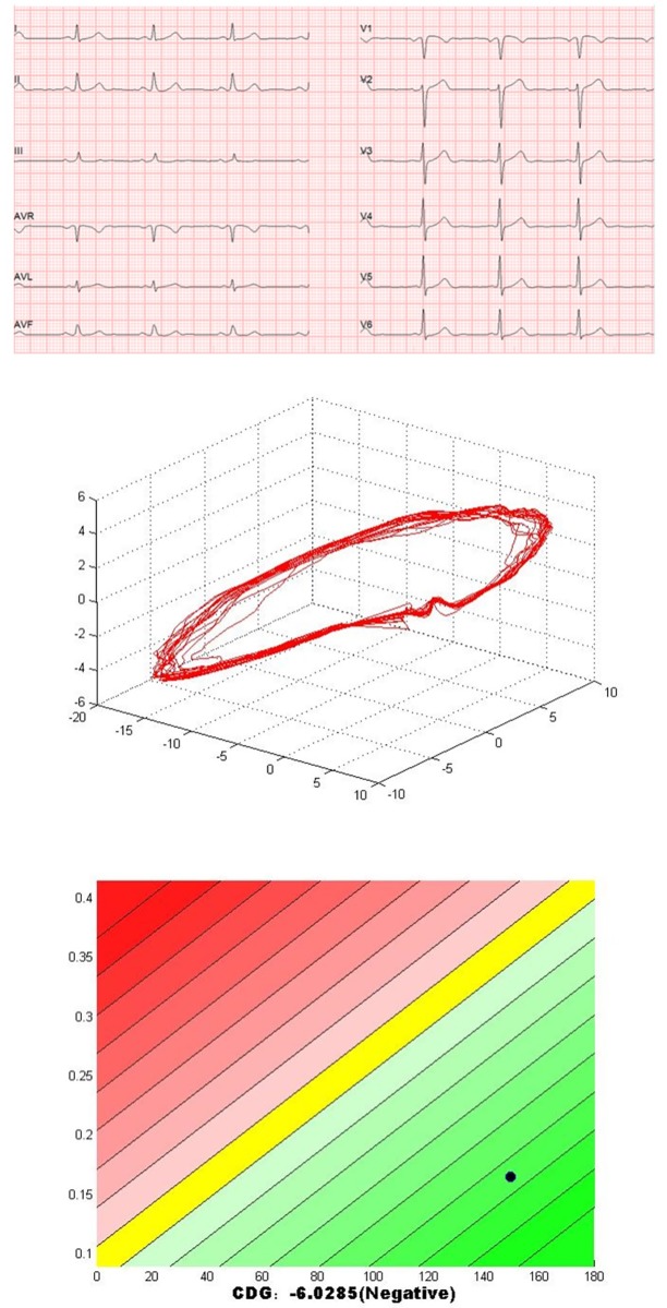
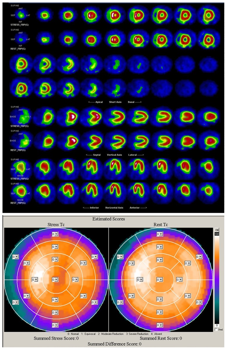
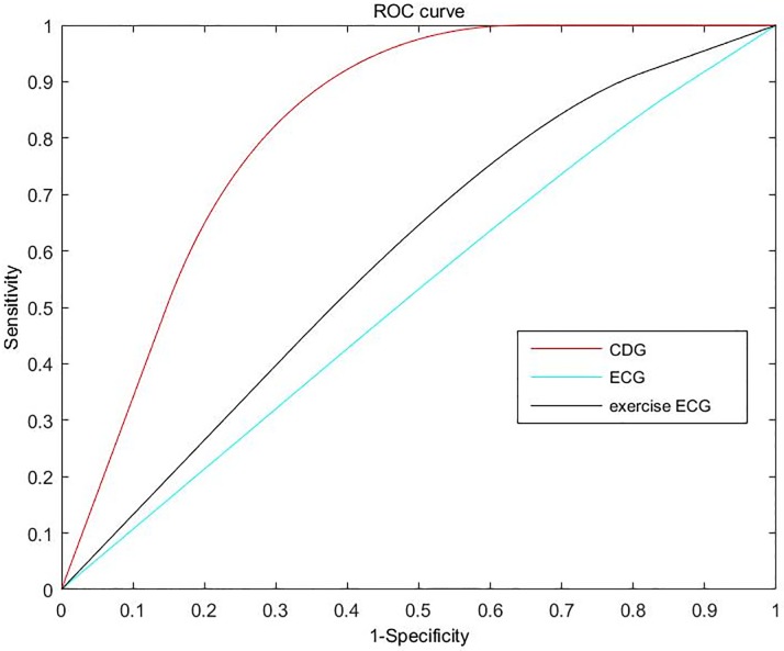
Myocardial perfusion abnormalities are the first sign of the ischemic cascade in the development of coronary artery disease (CAD). Thus, the early detection of myocardial perfusion abnormalities is significant for the prevention of CAD. Recently, a novel noninvasive method named Cardiodynamicsgram (CDG) has been proposed for early detection of CAD. This study aims to evaluate the predictive value of CDG in myocardial perfusion abnormalities for suspected ischemic heart disease. In the study, 86 suspected patients were enrolled. Standard 12-lead ECG and CDG were performed simultaneously before single-photon emission computed tomography (SPECT) myocardial perfusion imaging (MPI). Diagnostic accuracy of CDG for myocardial perfusion abnormalities detection is assessed using SPECT MPI as the reference standard. Of these 86 suspected patients, 37 patients were positive in CDG, 49 patients were negative in CDG. Diagnostic accuracy of CDG at presentation for myocardial perfusion abnormalities was 84.9%, sensitivity 84.0%, and specificity 89.4%. Furthermore, of the 10 patients whose SPECT MPI results are reverse redistribution, 9 patients were positive in CDG. Underlying causes of false positive CDG findings included the factors that can change the stability of cardiac electrical conduction and measurement noise. Myocardial remodeling in patients with old myocardial infarction might be the major cause of false negative findings. Results show a good consistency between the CDG and SPECT MPI in evaluating myocardial perfusion abnormalities. It suggests that CDG might be used as a cost-effective tool for assessing the myocardial perfusion abnormalities in the clinic.
Conflict of interest statement
The authors have declared that no competing interests exist.
Figures
References
-
- Organization WH. Cardiovascular diseases (CVDs); 2017. Available from: http://www.who.int/mediacentre/factsheets/fs317/en/.
-
- Qayyum A, Kastrup J. Measuring myocardial perfusion: the role of PET, MRI and CT. Clinical radiology. 2015;70(6):576–584. 10.1016/j.crad.2014.12.017 - DOI - PubMed
-
- Tung R, Bauer B, Schelbert H, Lynch JP III, Auerbach M, Gupta P, et al. Incidence of abnormal positron emission tomography in patients with unexplained cardiomyopathy and ventricular arrhythmias: the potential role of occult inflammation in arrhythmogenesis. Heart Rhythm. 2015;12(12):2488–2498. 10.1016/j.hrthm.2015.08.014 - DOI - PMC - PubMed
-
- Barizon GC, Simoes MV, Schmidt A, Gadioli LP, Murta LO Junior. Relationship between microvascular changes, autonomic denervation, and myocardial fibrosis in Chagas cardiomyopathy: Evaluation by MRI and SPECT imaging. Journal of Nuclear Cardiology. 2018; p. 1–11. - PubMed
-
- Beller GA. Future growth and success of nuclear cardiology. Journal of Nuclear Cardiology. 2018;25(2):375–378. 10.1007/s12350-018-1211-1 - DOI - PubMed
Publication types
MeSH terms
LinkOut - more resources
Full Text Sources
Medical
Miscellaneous
