Review
. 2019 Mar 1;6(1):R9-R23.
doi: 10.1530/ERP-18-0049.

EDUCATIONAL SERIES IN CONGENITAL HEART DISEASE: Tetralogy of Fallot: diagnosis to long-term follow-up

Affiliations
Review

EDUCATIONAL SERIES IN CONGENITAL HEART DISEASE: Tetralogy of Fallot: diagnosis to long-term follow-up

R Bedair et al. Echo Res Pract. .

Abstract

Tetralogy of Fallot (TOF) is the most common cyanotic congenital heart defect, affecting 3 in 10,000 live births. Surgical correction in early childhood is associated with good outcomes, but lifelong follow-up is necessary to identify the long-term sequelae that may occur. This article will cover the diagnosis of TOF in childhood, the objectives of surveillance through adulthood and the value of multi-modality imaging in identifying and guiding timely surgical and percutaneous interventions.

Keywords: congenital heart disease; multi-modality imaging; pulmonary regurgitation; tetralogy of Fallot.


Figures

Figure 1
Figure 1
(A) Classical tetralogy of Fallot with obstructive infundibular, valvular and pulmonary components. (B) The photograph, taken from the apex of the morphologically right ventricle looking towards the base, and the computed tomography of the anatomic model (C), show the phenotypic features of tetralogy of Fallot: antero-superior displacement of the conal septum causing moderate stenosis of the right ventricular outflow tract, and a VSD due to the misalignment of the outlet septum. The heart has been sectioned to replicate the oblique subcostal echocardiographic cut (Fig. 5). AoV, aortic valve; PV, pulmonary valve; RV, right ventricle; SB, septal band; SPT, septo parietal trabeculations; TV, tricuspid valve; VIF, ventriculo infundibular fold; VSD, ventrciular septal defect.
Figure 2
Figure 2
Coronary artery arrangement in patients with TOF. Parasternal short-axis echo image (A) and CT angiogram (B) showing an aberrant left main (LM) coronary artery arising from the right coronary artery (RCA) and crossing the RVOT, as well as a large conal branch in a patient with TOF. Ao, aorta; MPA, main pulmonary artery.
Figure 3
Figure 3
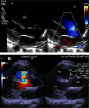
TOF with absent pulmonary valve and hypoplastic annulus: free pulmonary regurgitation (left panel); color Doppler flow acceleration in systole (right panel) and dilated MPA. MPA, main pulmonary artery.
Figure 4
Figure 4
Repaired TOF with absent pulmonary valve. Aneurysmal dilatation of the branch pulmonary arteries with the RPA causing right bronchial narrowing. LPA, left pulmonary artery; RPA, right pulmonary artery.
Figure 5
Figure 5
TOF-AVSD. Subcostal short-axis images at the level of the AV valve valve showing an en face view of the common AV valve (SBL, superior bridging leaflet, IBL, inferior bridging leaflet, LML, left mural leaflet are seen) and the deviation of the outlet septum.
Figure 6
Figure 6
Subcostal view. Subcostal short-axis view showing the overriding aorta and non-restrictive VSD with laminar flow on color flow mapping (A). Modified anterior oblique subcostal view shows the length of RVOT, pulmonary valve and branch pulmonary arteries in detail. Flow turbulence on color flow mapping confirms infundibular obstruction. Ao, aorta; LPA, left pulmonary artery; LV, let ventricle; RA, right atrium; RV, right ventricle; RVOT, right ventricular outflow tract; VSD, ventricular septal defect.
Figure 7
Figure 7
Parasternal long axis view. Parasternal long axis view showing a non-restrictive VSD (A) with right to left shunting and a restrictive VSD (B) with tricuspid leaflet tissue seen in the 2D imaging within the VSD. Ao, aorta; LV, left ventricle; RV, right ventricle; TV, tricuspid valve; VSD, ventricular septal defect.
Figure 8
Figure 8
Severe pulmonary regurgitation in a patient with repaired TOF. Parasternal short-axis window showing the main (MPA) and branch pulmonary arteries on 2D imaging (A) with pulmonary regurgitation on color flow mapping occupying the width of the MPA and originating from the two branches (B), with a short pressure half time (65 ms) on spectral Doppler imaging (C). LPA, left pulmonary artery; MPA, main pulmonary artery; RPA, right pulmonary artery.
Figure 9
Figure 9
Multi-level RV outflow tract assessment by CW Doppler. Spectral Doppler showing severe obstruction (peak velocity 5.2 m/s, peak gradient 108 mmHg) across RV infundibulum (Dagger shaped envelope) and pulmonary valve (symmetrical envelope). CW, continuous wave; RV, right ventricle.
Figure 10
Figure 10
Dilated right ventricle in a patient with repaired TOF and severe pulmonary regurgitation. Apical four chamber view in a patient with repaired TOF and severe pulmonary regurgitation before pulmonary valve replacement surgery, showing dilated right ventricle: RVD1 58 mm and right atrium, (A) and mildly reduced indices of basal longitudinal function, TAPSE 16 mm (B) and tricuspid annular TDI showing an S′ of 9 cm/s (C). RVD, right ventricle dimension; TAPSE, tricuspid annular systolic plane excursion; TDI, tissue Doppler imaging.
Figure 11
Figure 11
‘Restrictive RV’ physiology on spectral Doppler. Spectral Doppler across the pulmonary valve in a patient with previous TOF repair showing the characteristic ‘A’ wave (red arrow) in a patient with a restrictive RV. This pre-systolic forward flow in the pulmonary artery is caused by the pressure generated by right atrial contraction (dashed line indicating end of atrial contraction) transmitted through the right ventricle to the RVOT and causing pre-systolic opening of the pulmonary valve. RV, right ventricle; RVOT, right ventricular outflow tract.
Figure 12
Figure 12
Examples of scar distributions on high-resolution LGE images in patients with repaired TOF. LGE data from three patients are shown (A, B and C). Trans-axial LGE images are shown in the left column, yellow and green arrows indicating RVOT and septal scars, respectively. In the right column, the corresponding scar distributions are displayed in RVOT and septal views (left and right images, respectively). LGE, late gadolinium enhancement; RVOT, right ventricular outflow tract.
Figure 13
Figure 13
Right ventricular strain assessment. (A) Apical 4-chamber view with semi-automatic detection of the regions of interest. This method requires the user to outline the internal border of the myocardium. The 2D strain algorithm automatically evaluates the tracking quality at each myocardial location over time, and provides the tracking quality of each segment as either ‘acceptable’ (V) or ‘non-acceptable’ (X) in a table just below the image. (B) RV regional longitudinal peak systolic strain. (C) Myocardial longitudinal strain curves determined by speckle tracking imaging. (D) Map of the regional longitudinal strain.
Figure 14
Figure 14
3D RV assessment. (A) Display from the four-dimensional RV function analysis program showing the final stage of contour detection, in which manual correction of the contours can be applied in any cross-section or phase of the cardiac cycle. (B) View of the four-dimensional RV function mesh showing RV geometry and volume. RV, right ventricle.
Figure 15
Figure 15
Use of cardiac MRI to assess severity of pulmonary regurgitation using phase-contrast imaging. (A) Sagittal view of the right ventricular outflow tract. Line (red) indicating the plane of imaging for flow in the expected position of the pulmonary valve. (B) Circle indicating assessment of flow in region of interest. (C) Flow curves from phase-contrast imaging. Pulmonary regurgitation indicated flow below the baseline (arrow). Image courtesy of Dr Michael Yeong.
Figure 16
Figure 16
Cine SSFP short-axis ventricular stack for ventricular volumetric data. Dilated right ventricle indicated by arrow (white). Image courtesy of Dr Michael Yeong.
Figure 17
Figure 17
Assessment of suitability for percutaneous pulmonary valve implantation. Contrast cardiac CT (A) showing calcification in an RV–PA conduit (pulmonary homograft) in a patient with previous pulmonary atresia-VSD repair showing heavy calcification and position of the proximal left anterior descending (LAD) coronary artery in relation to the conduit indicated by the red arrow. Cardiac catheter with simultaneous balloon interrogation of the conduit and left coronary angiography (B) allows profiling of the conduit shows that the narrowest segment of the conduit (white arrow) is relatively distant from the LAD. LAD, left anterior descending coronary artery; LCx, left circumflex coronary artery; PA, pulmonary artery; RV, right ventricle.

References

    1. Loffredo CA. Epidemiology of cardiovascular malformations: prevalence and risk factors. American Journal of Medical Genetics 2000. 97 319–325. (10.1002/1096-8628(200024)97:4<319::AID-AJMG1283>3.0.CO;2-E) - DOI - PubMed
    1. Suzuki A, Ho SY, Anderson RH, Deanfield JE. Further morphologic studies on tetralogy of Fallot, with particular emphasis on the prevalence and structure of the membranous flap. Journal of Thoracic and Cardiovascular Surgery 1990. 99 528–535. - PubMed
    1. Ramaswamy P, Lytrivi ID, Thanjan MT, Nguyen T, Srivastava S, Sharma S, Ko HH, Parness IA, Lai WW. Frequency of aberrant subclavian artery, arch laterality, and associated intracardiac anomalies detected by echocardiography. American Journal of Cardiology 2008. 101 677–682. (10.1016/j.amjcard.2007.10.036) - DOI - PubMed
    1. Anderson RH, Weinberg PM. The clinical anatomy of tetralogy of fallot. Cardiology in the Young 2005. 15 (Supplement 1) 38–47. (10.1017/S1047951105001010) - DOI - PubMed
    1. Nakata S, Imai Y, Takanashi Y, Kurosawa H, Tezuka K, Nakazawa M, Ando M, Takao A. A new method for the quantitative standardization of cross-sectional areas of the pulmonary arteries in congenital heart diseases with decreased pulmonary blood flow. Journal of Thoracic and Cardiovascular Surgery 1984. 88 610–619. - PubMed

LinkOut - more resources