

CT-pathologic correlation in lung adenocarcinoma and squamous cell carcinoma
- PMID: 30557988
- PMCID: PMC6320064
- DOI: 10.1097/MD.0000000000013362
CT-pathologic correlation in lung adenocarcinoma and squamous cell carcinoma
Abstract
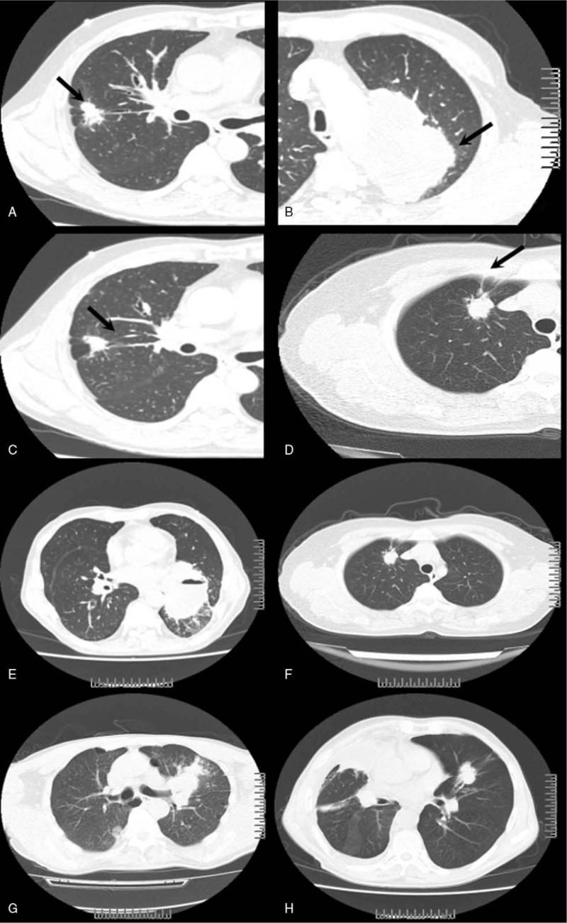
Distinguishing lung adenocarcinoma from squamous cell carcinoma (SCC) is clinically important. Computed tomography (CT) scan is an economical, effective, noninvasive, commonly available, and quick diagnostic way for lung cancer. In this study, we aim to compare the CT characteristics in adenocarcinoma and SCC.Data from 275 cases (259 adenocarcinoma and 16 SCC) were retrospectively compared. CT characteristics, including lesion size and shape, single/multifocal lesions, location of the tumor, the margin of lobes, whether the lesion had deep lobulated margin, bronchial cut-off sign, signs of dilated bronchial arteries, signs of vascular bundle thickening, signs of short burrs, spinous processes, and pleural indentation, were compared in 148 cases (137 adenocarcinoma and 11 SCC).Patients with adenocarcinoma were more likely to be female (44.2% vs 25.0%, P = .017). Compared with SCC, adenocarcinomas were more likely to have deep lobulated margin (81.0% vs 54.5%, P = .038), less likely to have smooth lobes margin (2.7% vs 83.3%, P < .001), more likely to have vascular bundle thickening (37.2% vs 0, P = .016) and pleural indentation (59.9% vs 18.2%, P = .01), and marginally less likely to have dilated bronchial arteries (17.5% vs 45.5%, P = .064). No significant difference was observed regarding to characteristics, including tumor size, location of the tumor, signs of bronchial cut-off, dilated bronchial arteries, short burrs, or spinous processes.CT scan has the potential to help to distinguish lung adenocarcinoma and SCC in a fast and commonly available way. CT could be a rough but fast way to diagnosis, and may thus shorten the waiting time to treatment and allow more time for clinicians, patients, and their families to prepare for future treatment.
Conflict of interest statement
The author(s) of this work have nothing to disclose and have no conflicts of interest.
Figures
References
-
- Siegel RL, Miller KD, Jemal A. Cancer statistics, 2015. CA Cancer J Clin 2015;65:5–29. - PubMed
-
- Asamura H, Goya T, Koshiishi Y, et al. A Japanese Lung Cancer Registry study: prognosis of 13,010 resected lung cancers. J Thorac Oncol 2008;3:46–52. - PubMed
-
- Kim HL, Puymon MR, Qin M, et al. NCCN clinical practice guidelines in oncology™, 2013.
-
- Shigematsu H, Lin L, Takahashi T, et al. Clinical and biological features associated with epidermal growth factor receptor gene mutations in lung cancers. J Natl Cancer Inst 2005;97:339–46. - PubMed
-
- Scagliotti G, Brodowicz T, Shepherd FA, et al. Treatment-by-histology interaction analyses in three phase III trials show superiority of pemetrexed in nonsquamous non-small cell lung cancer. J Thorac Oncol 2011;6:64–70. - PubMed
MeSH terms
LinkOut - more resources
Full Text Sources
Medical
Research Materials
