

Perspectives of RAS and RHEB GTPase Signaling Pathways in Regenerating Brain Neurons
- PMID: 30558189
- PMCID: PMC6321366
- DOI: 10.3390/ijms19124052
Perspectives of RAS and RHEB GTPase Signaling Pathways in Regenerating Brain Neurons
Abstract
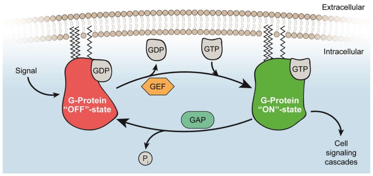
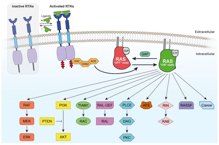
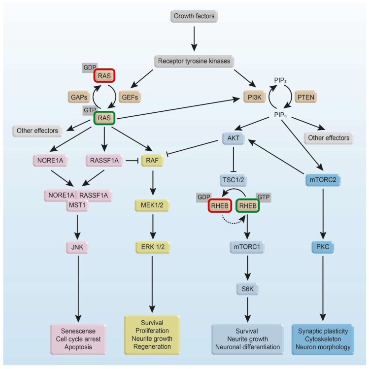
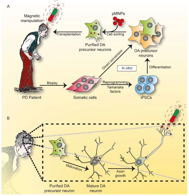
Cellular activation of RAS GTPases into the GTP-binding "ON" state is a key switch for regulating brain functions. Molecular protein structural elements of rat sarcoma (RAS) and RAS homolog protein enriched in brain (RHEB) GTPases involved in this switch are discussed including their subcellular membrane localization for triggering specific signaling pathways resulting in regulation of synaptic connectivity, axonal growth, differentiation, migration, cytoskeletal dynamics, neural protection, and apoptosis. A beneficial role of neuronal H-RAS activity is suggested from cellular and animal models of neurodegenerative diseases. Recent experiments on optogenetic regulation offer insights into the spatiotemporal aspects controlling RAS/mitogen activated protein kinase (MAPK) or phosphoinositide-3 kinase (PI3K) pathways. As optogenetic manipulation of cellular signaling in deep brain regions critically requires penetration of light through large distances of absorbing tissue, we discuss magnetic guidance of re-growing axons as a complementary approach. In Parkinson's disease, dopaminergic neuronal cell bodies degenerate in the substantia nigra. Current human trials of stem cell-derived dopaminergic neurons must take into account the inability of neuronal axons navigating over a large distance from the grafted site into striatal target regions. Grafting dopaminergic precursor neurons directly into the degenerating substantia nigra is discussed as a novel concept aiming to guide axonal growth by activating GTPase signaling through protein-functionalized intracellular magnetic nanoparticles responding to external magnets.
Keywords: Magneto Protein Therapy; Parkinson’s disease; RAS GTPase; RHEB GTPase; axonal guidance; brain regeneration; magnetogenetics; nanoparticle; optogenetics; survival.
Conflict of interest statement
The authors declare no conflict of interest.
Figures
Similar articles
-
Novel Tools towards Magnetic Guidance of Neurite Growth: (I) Guidance of Magnetic Nanoparticles into Neurite Extensions of Induced Human Neurons and In Vitro Functionalization with RAS Regulating Proteins.J Funct Biomater. 2019 Jul 16;10(3):32. doi: 10.3390/jfb10030032. J Funct Biomater. 2019. PMID: 31315182 Free PMC article.
-
Protection of nigral dopaminergic neurons by AAV1 transduction with Rheb(S16H) against neurotoxic inflammation in vivo.Exp Mol Med. 2018 Feb 9;50(2):e440. doi: 10.1038/emm.2017.261. Exp Mol Med. 2018. PMID: 29422542 Free PMC article.
-
Rit subfamily small GTPases: regulators in neuronal differentiation and survival.Cell Signal. 2013 Oct;25(10):2060-8. doi: 10.1016/j.cellsig.2013.06.002. Epub 2013 Jun 11. Cell Signal. 2013. PMID: 23770287 Free PMC article. Review.
-
Upregulation of Neuronal Rheb(S16H) for Hippocampal Protection in the Adult Brain.Int J Mol Sci. 2020 Mar 16;21(6):2023. doi: 10.3390/ijms21062023. Int J Mol Sci. 2020. PMID: 32188096 Free PMC article. Review.
-
Coordination of Rheb lysosomal membrane interactions with mTORC1 activation.F1000Res. 2020 May 27;9:F1000 Faculty Rev-450. doi: 10.12688/f1000research.22367.1. eCollection 2020. F1000Res. 2020. PMID: 32518628 Free PMC article. Review.
Cited by
-
Galectin-1 confers resistance to doxorubicin in hepatocellular carcinoma cells through modulation of P-glycoprotein expression.Cell Death Dis. 2022 Jan 24;13(1):79. doi: 10.1038/s41419-022-04520-6. Cell Death Dis. 2022. PMID: 35075112 Free PMC article.
-
β-Arrestin2 Is Critically Involved in the Differential Regulation of Phosphosignaling Pathways by Thyrotropin-Releasing Hormone and Taltirelin.Cells. 2022 Apr 27;11(9):1473. doi: 10.3390/cells11091473. Cells. 2022. PMID: 35563779 Free PMC article.
-
Novel Tools towards Magnetic Guidance of Neurite Growth: (I) Guidance of Magnetic Nanoparticles into Neurite Extensions of Induced Human Neurons and In Vitro Functionalization with RAS Regulating Proteins.J Funct Biomater. 2019 Jul 16;10(3):32. doi: 10.3390/jfb10030032. J Funct Biomater. 2019. PMID: 31315182 Free PMC article.
-
siRNA Mediated Downregulation of RhoA Expression Reduces Oxidative Induced Apoptosis in Retinal Ganglion Cells.Curr Mol Med. 2024;24(5):630-636. doi: 10.2174/1566524023666230511095628. Curr Mol Med. 2024. PMID: 37171014
-
Parallelized Manipulation of Adherent Living Cells by Magnetic Nanoparticles-Mediated Forces.Int J Mol Sci. 2020 Sep 8;21(18):6560. doi: 10.3390/ijms21186560. Int J Mol Sci. 2020. PMID: 32911745 Free PMC article.
References
-
- Kirsten W.H., Mayer L.A. Morphologic responses to a murine erythroblastosis virus. J. Natl. Cancer Inst. 1967;39:311–335. - PubMed
Publication types
MeSH terms
Substances
Grants and funding
LinkOut - more resources
Full Text Sources
Research Materials
Miscellaneous
