. 2018 Dec 16;7(12):273.
doi: 10.3390/cells7120273.

Tangential Flow Filtration for Highly Efficient Concentration of Extracellular Vesicles from Large Volumes of Fluid

Affiliations

Tangential Flow Filtration for Highly Efficient Concentration of Extracellular Vesicles from Large Volumes of Fluid

Sara Busatto et al. Cells. .

Abstract

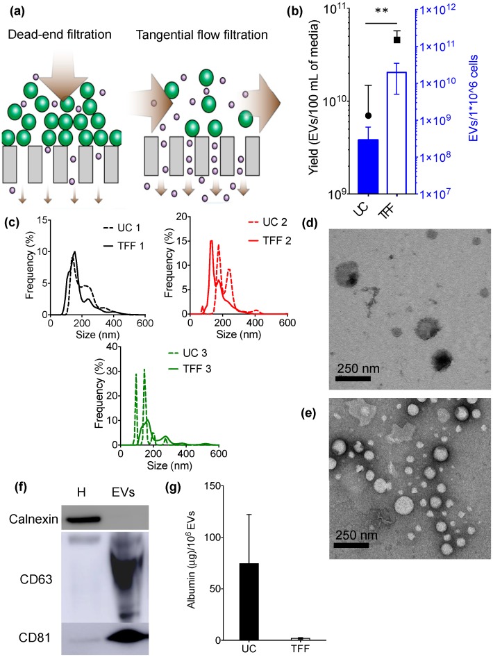
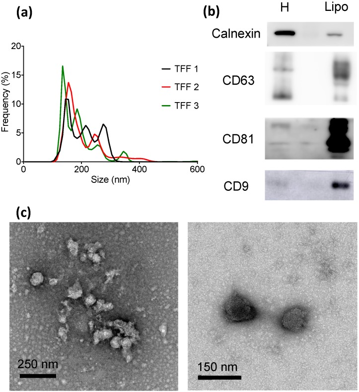
Concentration of extracellular vesicles (EVs) from biological fluids in a scalable and reproducible manner represents a major challenge. This study reports the use of tangential flow filtration (TFF) for the highly efficient isolation of EVs from large volumes of samples. When compared to ultracentrifugation (UC), which is the most widely used method to concentrate EVs, TFF is a more efficient, scalable, and gentler method. Comparative assessment of TFF and UC of conditioned cell culture media revealed that the former concentrates EVs of comparable physicochemical characteristics, but with higher yield, less single macromolecules and aggregates (<15 nm in size), and improved batch-to-batch consistency in half the processing time (1 h). The TFF protocol was then successfully implemented on fluids derived from patient lipoaspirate. EVs from adipose tissue are of high clinical relevance, as they are expected to mirror the regenerative properties of the parent cells.

Keywords: exosome; extracellular vesicles; isolation; lipoaspirate; tangential flow filtration; ultrafiltration.


Conflict of interest statement

The authors declare no conflict of interest.

Figures

Figure 1
Figure 1
Biophysical and biochemical characterization of extracellular vesicles (EVs) isolated from MDA-MB-231 breast cancer cell culture media using ultracentrifugation (UC) or tangential flow filtration (TFF). (a) Schematic illustrating the difference between dead-end filtration and TFF; (b) EV yield; (c) Size distribution (0–650 nm) of EVs isolated with UC (dashed lines) and TFF (continuous lines); (d,e) Transmission electron microscopy (TEM) of EVs obtained with UC (d) or TFF (e); (f) Western blot of characteristic intracellular (calnexin) and EV (CD63, CD81) markers in samples isolated by TFF; (g) Levels of albumin contaminants in EV samples. Cellular homogenate (H) was used as a control. Data are presented as mean ± s.d. of three biological replicates (b,c) or experimental replicates (g). Statistical significance was evaluated by Student’s t-test. ** p < 0.01.
Figure 2
Figure 2
Biophysical and biochemical characterization of EV-enriched lipoaspirate-derived samples (Lipo) isolated with TFF; (a) Size distribution of EVs (triplicate); (b) Western blot of characteristic intracellular (calnexin) and EV (CD63, CD81, CD9) markers. Cellular homogenate (H) was used as a control; (c) TEM images of EV-enriched formulations.

References

    1. Colombo M., Raposo G., Thery C. Biogenesis, secretion, and intercellular interactions of exosomes and other extracellular vesicles. Annu. Rev. Cell Dev. Biol. 2014;30:255–289. doi: 10.1146/annurev-cellbio-101512-122326. - DOI - PubMed
    1. Candelario K.M., Steindler D.A. The role of extracellular vesicles in the progression of neurodegenerative disease and cancer. Trends Mol. Med. 2014;20:368–374. doi: 10.1016/j.molmed.2014.04.003. - DOI - PMC - PubMed
    1. Vader P., Breakefield X.O., Wood M.J. Extracellular vesicles: Emerging targets for cancer therapy. Trends Mol. Med. 2014;20:385–393. doi: 10.1016/j.molmed.2014.03.002. - DOI - PMC - PubMed
    1. Borrelli D.A., Yankson K., Shukla N., Vilanilam G., Ticer T., Wolfram J. Extracellular vesicle therapeutics for liver disease. J. Control. Release. 2018;273:86–98. doi: 10.1016/j.jconrel.2018.01.022. - DOI - PubMed
    1. Wu Y., Deng W., Klinke D.J., 2nd Exosomes: Improved methods to characterize their morphology, RNA content, and surface protein biomarkers. Analyst. 2015;140:6631–6642. doi: 10.1039/C5AN00688K. - DOI - PMC - PubMed