

Juvenile open-angle Glaucoma associated with Leber's hereditary optic neuropathy: a case report and literature review
- PMID: 30558558
- PMCID: PMC6296145
- DOI: 10.1186/s12886-018-0980-2
Juvenile open-angle Glaucoma associated with Leber's hereditary optic neuropathy: a case report and literature review
Abstract
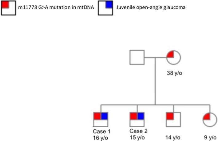
Background: Leber's hereditary optic neuropathy (LHON) is a maternally inherited recessive disease rarely complicated with glaucoma. We conducted a clinical and genetic retrospective case series to describe three cases of juvenile open-angle glaucoma (JOAG) and an ND4 m11778G > A mitochondrial DNA (mtDNA) mutation, which is pathognomonic for LHON.
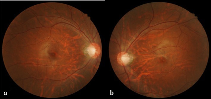
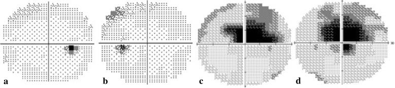
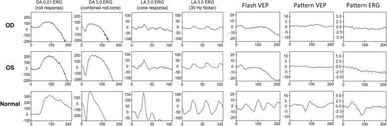
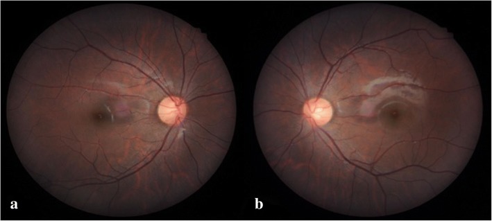
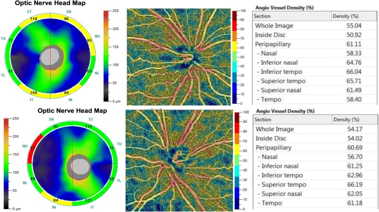
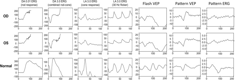
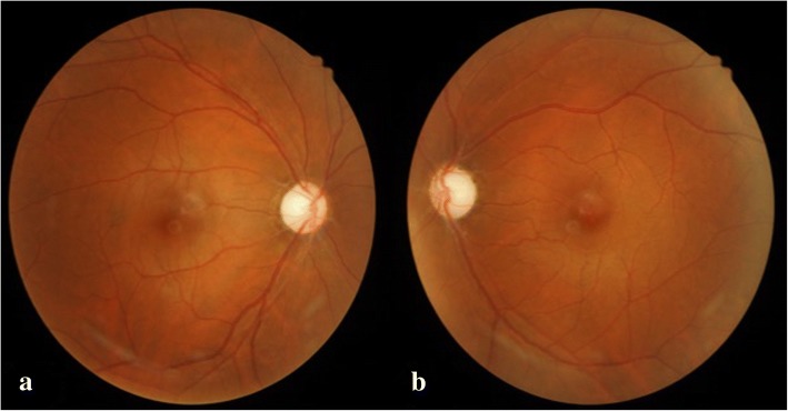
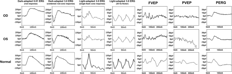
Case presentation: Patient 1 was a 16-year-old boy diagnosed with bilateral JOAG and high myopia. His intraocular pressure (IOP) was poorly controlled with the use of full topical anti-glaucoma medications. His best-corrected visual acuity (BCVA) decreased gradually over 5 years. Fundoscopic examination revealed bilateral enlarged disc cupping of the optic nerves with sectorial excavation and reduction of the neural rim in the left eye. His visual field (VF) was characterized by bilateral progressive central scotoma. Pattern visual evoked potentials (VEPs) and pattern electroretinograms (ERGs) showed extinguished responses in both eyes. Because of the non-specific visual field findings and the optic neuropathy disclosed by the pattern VEPs and pattern ERGs, we arranged a genetic test for the patient, which revealed an m11778G > A mtDNA mutation. Patient 2, the younger brother of Patient 1, was a 15-year-old boy who had been diagnosed with bilateral JOAG in 2010. The BCVA of both eyes remained at 1.0 during the follow-up period. Fundoscopic examination revealed bilateral mildly paled optic disc with enlarged cupping and reduction of the neural rim. The pattern ERG revealed a decreased N95 amplitude bilaterally. The genetic test revealed an m11778G > A mtDNA mutation. Patient 3 was a 35-year-old man with bilateral JOAG. His BCVA decreased gradually over 10 years. Fundoscopic examination revealed paled optic disc with enlarged disc cupping and reduction of the neural rim in both eyes. The pattern ERG revealed a decreased N95 amplitude bilaterally. The genetic test revealed an m11778G > A mtDNA mutation.
Conclusions: This case series describes three patients with concomitant occurrence of JOAG and LHON. These two diseases may have a cumulative effect on oxidative stress and retinal ganglion cell death with the rapid deterioration of vision, which may occur during adolescence.
Keywords: Juvenile open-angle glaucoma; Leber’s hereditary optic neuropathy; Mitochondria.
Conflict of interest statement
Ethics approval and consent to participate
This study was approved by the Institutional Review Board of Chang Gung Medical Foundation. IRB No.: 201700707B0.
Consent for publication
Consent to publish has been obtained from the patients or their parents.
Competing interests
The authors declare that they have no competing interests.
Publisher’s Note
Springer Nature remains neutral with regard to jurisdictional claims in published maps and institutional affiliations.
Figures
Similar articles
-
Glaucoma progression associated with Leber's hereditary optic neuropathy.Int Ophthalmol. 2013 Feb;33(1):75-7. doi: 10.1007/s10792-012-9623-4. Epub 2012 Sep 17. Int Ophthalmol. 2013. PMID: 22983441
-
Leber's hereditary optic neuropathy mitochondrial DNA mutations in normal-tension glaucoma.Graefes Arch Clin Exp Ophthalmol. 2001 Jul;239(6):437-40. doi: 10.1007/s004170100309. Graefes Arch Clin Exp Ophthalmol. 2001. PMID: 11561792
-
[Past, present, and future in Leber's hereditary optic neuropathy].Nippon Ganka Gakkai Zasshi. 2001 Dec;105(12):809-27. Nippon Ganka Gakkai Zasshi. 2001. PMID: 11802455 Review. Japanese.
-
[Rapid onset of visual recovery following acute visual loss due to leber's hereditary optic neuropathy].Rev Neurol (Paris). 2005 May;161(5):599-601. doi: 10.1016/s0035-3787(05)85099-4. Rev Neurol (Paris). 2005. PMID: 16106816 French.
-
[DNA diagnosis in the age of individual made-to-order medications].Nippon Ganka Gakkai Zasshi. 2004 Dec;108(12):863-85; discussion 886. Nippon Ganka Gakkai Zasshi. 2004. PMID: 15656090 Review. Japanese.
Cited by
-
Leber's hereditary optic neuropathy: Update on current diagnosis and treatment.Front Ophthalmol (Lausanne). 2023 Jan 11;2:1077395. doi: 10.3389/fopht.2022.1077395. eCollection 2022. Front Ophthalmol (Lausanne). 2023. PMID: 38983564 Free PMC article. Review.
-
Extranuclear DNA Variations in the Susceptibility of Glaucoma.Middle East Afr J Ophthalmol. 2024 Jun 14;30(2):113-120. doi: 10.4103/meajo.meajo_132_23. eCollection 2023 Apr-Jun. Middle East Afr J Ophthalmol. 2024. PMID: 39006929 Free PMC article. Review.
-
Mitochondrial DNA variants in a cohort from Argentina with suspected Leber's hereditary optic neuropathy (LHON).PLoS One. 2023 Feb 24;18(2):e0275703. doi: 10.1371/journal.pone.0275703. eCollection 2023. PLoS One. 2023. PMID: 36827238 Free PMC article.
-
Optic atrophy-associated TMEM126A is an assembly factor for the ND4-module of mitochondrial complex I.Proc Natl Acad Sci U S A. 2021 Apr 27;118(17):e2019665118. doi: 10.1073/pnas.2019665118. Proc Natl Acad Sci U S A. 2021. PMID: 33879611 Free PMC article.
-
Eye involvement in inherited metabolic disorders.Ther Adv Ophthalmol. 2020 Dec 29;12:2515841420979109. doi: 10.1177/2515841420979109. eCollection 2020 Jan-Dec. Ther Adv Ophthalmol. 2020. PMID: 33447730 Free PMC article. Review.
References
-
- Wallace DC. A new manifestation of Leber's disease and a new explanation for the agency responsible for its unusual pattern of inheritance. Brain. 1970;93(1):121–132. - PubMed
-
- Wallace DC, Singh G, Lott MT, Hodge JA, Schurr TG, Lezza AM, et al. Mitochondrial DNA mutation associated with Leber's hereditary optic neuropathy. Science. 1988;242(4884):1427–1430. - PubMed
Publication types
MeSH terms
Substances
LinkOut - more resources
Full Text Sources
