. 2018 Dec 17;15(1):78.
doi: 10.1186/s12977-018-0459-5.

Natural APOBEC3C variants can elicit differential HIV-1 restriction activity

Affiliations

Natural APOBEC3C variants can elicit differential HIV-1 restriction activity

Brett D Anderson et al. Retrovirology. .

Abstract

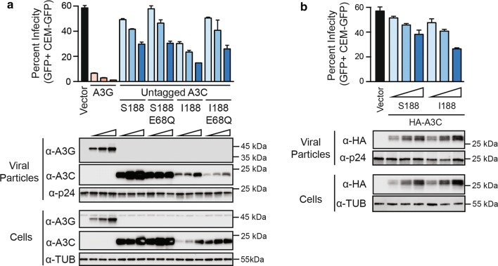
Background: The APOBEC3 (A3) family of DNA cytosine deaminases provides an innate barrier to infection by retroviruses including HIV-1. A total of five enzymes, A3C, A3D, A3F, A3G and A3H, are degraded by the viral accessory protein Vif and expressed at high levels in CD4+ T cells, the primary reservoir for HIV-1 replication in vivo. Apart from A3C, all of these enzymes mediate restriction of Vif-deficient HIV-1. However, a rare variant of human A3C (Ile188) was shown recently to restrict Vif-deficient HIV-1 in a 293T-based single cycle infection system. The potential activity of this naturally occurring A3C variant has yet to be characterized in a T cell-based spreading infection system. Here we employ a combination of Cas9/gRNA disruption and transient and stable protein expression to assess the roles of major Ser188 and minor Ile188 A3C variants in HIV-1 restriction in T cell lines.

Results: Cas9-mediated mutation of endogenous A3C in the non-permissive CEM2n T cell line did not alter HIV-1 replication kinetics, and complementation with A3C-Ser188 or A3C-Ile188 was similarly aphenotypic. Stable expression of A3C-Ser188 in the permissive T cell line SupT11 also had little effect. However, stable expression of A3C-Ile188 in SupT11 cells inhibited Vif-deficient virus replication and inflicted G-to-A mutations.

Conclusions: A3C-Ile188 is capable of inhibiting Vif-deficient HIV-1 replication in T cells. Although A3C is eclipsed by the dominant anti-viral activities of other A3s in non-permissive T cell lines and primary T lymphocytes, this enzyme may still be able to contribute to HIV-1 diversification in vivo. Our results highlight the functional redundancy in the human A3 family with regards to HIV-1 restriction and the need to consider naturally occurring variants.

Keywords: APOBEC3C; DNA cytosine deaminase; HIV-1; Human genetic variation; Innate immunity; Retrovirus restriction factor.


Figures

Fig. 1
Fig. 1
Expression of epitope-tagged and untagged A3C derivative constructs in 293T cells. Immunoblots of 293T cells transfected with 100 ng or 400 ng of C-terminally tagged (A3C-HA), N-terminally tagged (HA-A3C), or untagged A3C variants using either an anti-HA or anti-A3C antibody for detection. Tubulin was used as a loading control
Fig. 2
Fig. 2
A3C-Ile188 exhibits enhanced HIV-1 restriction activity in 293T cells. a Single cycle infectivity data for Vif-deficient HIV-1 viruses produced in the presence of untagged A3C-S188, A3C-I188, or catalytic mutant derivatives (E68Q). Immunoblots are shown below for viral particles (anti-A3G, anti-A3C, and anti-p24) and producer cells (anti-A3G, anti-A3C, and anti-tubulin). b Single cycle infectivity data for Vif-deficient HIV-1 viruses produced in the presence of N-terminally HA-tagged A3C-S188 or A3C-I188. Immunoblots are shown below for viral particles (anti-HA and anti-p24) and producer cells (anti-HA and anti-tubulin). All single cycle experiments were repeated at least 3 times, with representative infectivity data (mean ± SD) and immunoblots shown for one experiment
Fig. 3
Fig. 3
HIV-1 replication phenotypes following A3C disruption and variant complementation in non-permissive CEM2n cells. a Immunoblots of endogenous A3C in SupT11, H9, and CEM2n cells. Tubulin was used as a loading control. b Immunoblots of endogenous A3C in CEM2n clones following targeted disruption of A3C exon 3 by Cas9/gRNA complexes. Tubulin was used as a loading control. c A3C exon 3 sequences from parental CEM2n and an A3C-disrupted clone (CEM2n ∆A3C). d Flow cytometry plots for CEM2n ΔA3C cell pools 72 h post-transduction with GFP-reporter complementation vectors. The percentage of GFP+ cells is indicated for each population. e Immunoblots of A3C in the parental CEM2n line, CEM2n ΔA3C, and complemented CEM2n ΔA3C derivatives. Tubulin was used as a loading control. f, g Spreading infection kinetics of Vif-proficient and Vif-deficient HIV-1 (initial MOI = 0.02) in CEM2n, CEM2n ΔA3C, and the indicated complemented conditions. SupT11 cells are included as a control permissive cell type. Virus infectivity was determined by infection of CEM-GFP with culture supernatants followed 48 h later by quantification with flow cytometry. Each spreading infection experiment was performed 3 times and representative data are shown
Fig. 4
Fig. 4
Stable Expression of both A3C-Ile188 and A3C-Ser188 in SupT11 cells provides a partial block to Vif-deficient HIV-1 replication. a Flow cytometry plots for SupT11 cells 72 h after transduction with the indicated GFP-marked complementation vectors. The percentage of GFP+ cells is indicated for each condition. b Immunoblots of A3C in SupT11 cells transduced with constructs expressing A3C-Ser188, A3C-Ile188, or an empty control vector. Tubulin was used as a loading control. c Representative spreading infection data for Vif-deficient HIV-1 (MOI = 0.01) in SupT11 cells expressing A3C-Ser188, A3C-Ile188, or an empty control vector. Virus infectivity was determined by infection of CEM-GFP with culture supernatants followed 48 h later by quantification with flow cytometry. These data are from one of four biologically independent experiments. d Average G-to-A mutation loads for each condition. Error bars show ± SD of 4 independent experiments. Statistical comparisons were done using Student’s t test. p values above each panel are in comparison to vector control or A3C-Ser188. e Mutation data for each condition

References

    1. Altfeld M, Gale M., Jr Innate immunity against HIV-1 infection. Nat Immunol. 2015;16:554–562. doi: 10.1038/ni.3157. - DOI - PubMed
    1. Simon V, Bloch N, Landau NR. Intrinsic host restrictions to HIV-1 and mechanisms of viral escape. Nat Immunol. 2015;16:546–553. doi: 10.1038/ni.3156. - DOI - PMC - PubMed
    1. Harris RS, Hultquist JF, Evans DT. The restriction factors of human immunodeficiency virus. J Biol Chem. 2012;287:40875–40883. doi: 10.1074/jbc.R112.416925. - DOI - PMC - PubMed
    1. Harris RS, Dudley JP. APOBECs and virus restriction. Virology. 2015;479-480C:131–145. doi: 10.1016/j.virol.2015.03.012. - DOI - PMC - PubMed
    1. Stavrou S, Ross SR. APOBEC3 proteins in viral immunity. J Immunol. 2015;195:4565–4570. doi: 10.4049/jimmunol.1501504. - DOI - PMC - PubMed

Publication types

Substances