. 2018 Dec 17;18(1):1257.
doi: 10.1186/s12885-018-5096-9.

Expression of FGD4 positively correlates with the aggressive phenotype of prostate cancer

Affiliations

Expression of FGD4 positively correlates with the aggressive phenotype of prostate cancer

Alexia Bossan et al. BMC Cancer. .

Abstract

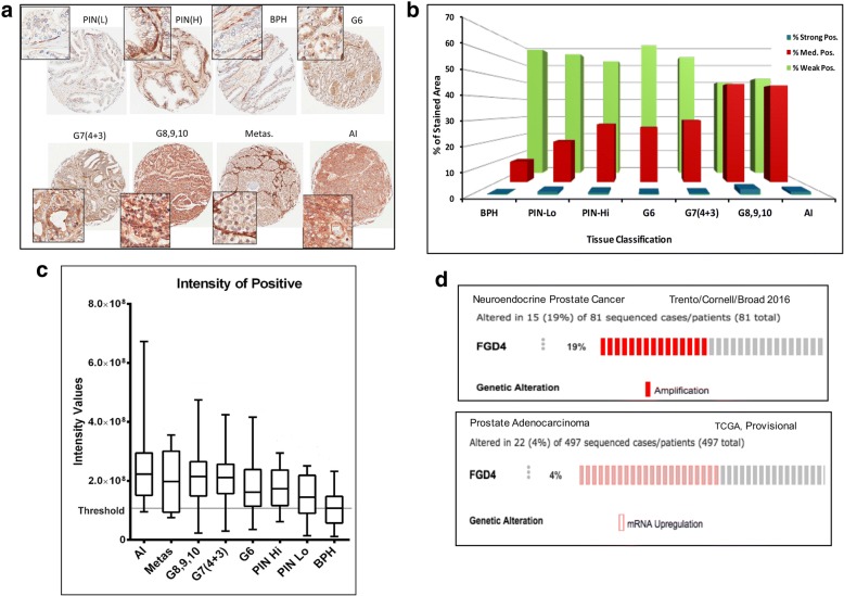
Background: FGD4 (Frabin) is an F-actin binding protein with GTP/GDP exchange activity specific for CDC42. It is involved in reorganization of the actin cytoskeleton, which requires both actin binding and CDC42 activating function of FGD4. Expression of FGD4 is altered in patients with heterogeneous hereditary motor and sensory neuropathies as a result of demyelination of peripheral nerves.

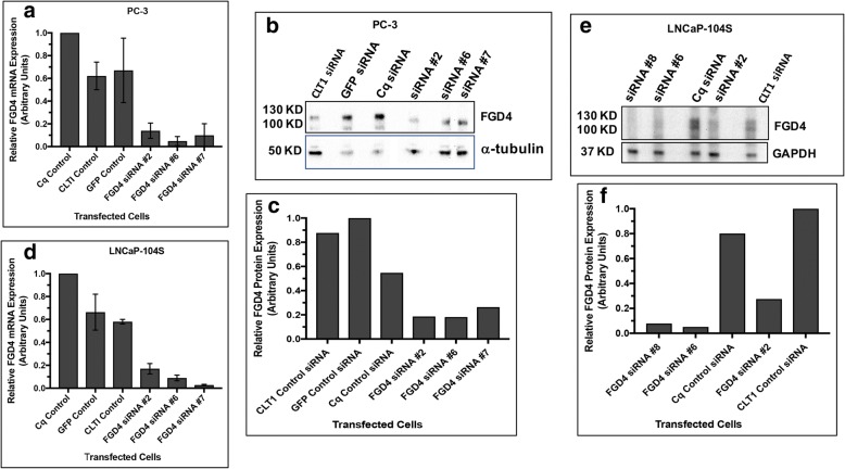
Methods: In this study, we examined the expression of FGD4 in prostate cancer specimens using immunohistochemistry and studied the function of FGD4 in maintaining cell phenotype, behavior and drug sensitivity using overexpression and siRNA-based silencing approaches. We used Mann-Whitney test for comparative analysis of FGD4 expression.

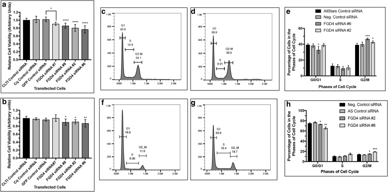
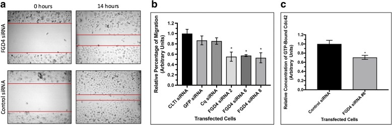
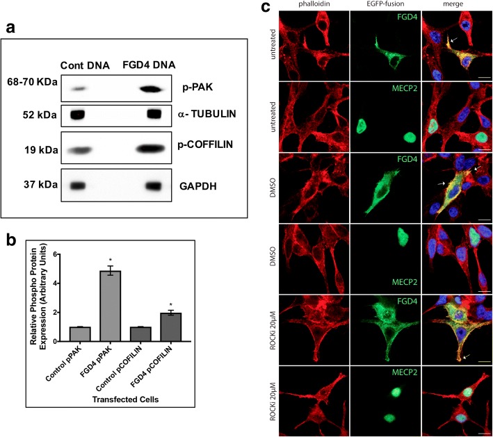
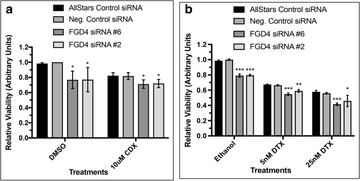
Results: Our results show that the expression of FGD4 is upregulated in cancerous prostates compared to the luminal cells in benign prostatic hyperplasia, although the basal cells showed high staining intensities. We noted a gradual increase in the staining intensity of FGD4 with increasing aggressiveness of the disease. Inhibition of expression of FGD4 using siRNAs showed reduced proliferation and cell cycle arrest in G2/M phase of androgen dependent LNCaP-104S and androgen refractory PC-3 cells. Inhibition of FGD4 also resulted in reduced cell migration and CDC42 activities in PC-3 cells whereas, ectopic expression of FGD4 induced cell migration, altered expression of mesenchymal and epithelial markers and activation of CDC42/PAK signaling pathway. Reduced expression of FGD4 improved sensitivity of LNCaP-104S cells to the anti-androgen drug Casodex and PC-3 cells to the microtubule stabilizing drug docetaxel.

Conclusions: Our data demonstrate a tumor promoting and a cell migratory function of FGD4 in prostate cancer cells and that inhibition of FGD4 expression enhances the response for both androgen-dependent and independent prostate cancer cells towards currently used prostate cancer drugs.

Keywords: Cell cycle; Cell migration; Drug sensitivity; Prostate cancer; Rho GEF.


Conflict of interest statement

Ethics approval and consent to participate

The prostate TMA construction was approved by the Moffitt scientific review committee (Reference # MCC13579) and by the USF’s institutional review board (IRB # USF 101642).

Consent for publication

Not applicable.

Competing interests

The authors declare that they have no competing interests.

Publisher’s Note

Springer Nature remains neutral with regard to jurisdictional claims in published maps and institutional affiliations.

Figures

Fig. 1
Fig. 1
Expression of FGD4 in prostate tumors changes with disease progression. a: Representative TMA images of the immunohistochemical analysis of FGD4 expression in benign prostatic hyperplasia (BPH) and prostate tumor tissues of different Gleason Grades, metastatic stages, and castration resistant (AI) stages. Images show weak staining in the basal cells in PIN (L) while strong staining in BPH, PIN (HI) tissues. A moderately higher expression of FGD4 could be noted in the luminal cells of prostate tumors with Gleason Scores 6 and 7, whereas a strong expression is evident in prostate tumor exhibiting more advanced stages. b: Comparative analysis of the percentage of strong, medium and low positive staining areas in BPH and prostate cancer tissues. c: Comparative analysis of the staining intensity across different prostate tissues. d: TCGA database analysis showing alteration in FGD4 DNA copy number and mRNA expression in neuroendocrine prostate cancer and prostate adenocarcinoma.
Fig. 2
Fig. 2
Inhibition of FGD4 expression upon FGD4 siRNA transfection of prostate cancer cells. a and d: Quantitative real-time PCR analysis of FGD4 mRNA in siRNA or control RNA transfected PC-3 and LNCaP-104S cells. Raw data have been normalized to the average of RPL13A and GAPDH, two control genes. Data show at least 70% reduction in FGD4 mRNA by FGD4 siRNAs compared to control siRNAs. b and e: Immunoblot analysis of PC-3 and LNCaP-104S cell lysates transfected with FGD4 siRNAs showing substantial reduction in FGD4 upon siRNA transfection. Alpha-tubulin and GAPDH were used as the loading controls c and f: Densitometric analyses of protein concentration normalized to the internal controls.
Fig. 3
Fig. 3
Inhibition of FGD4 decreased cell proliferation and arrested cells in G2/M phase. Cell proliferation assay showing a significant reduction in proliferation of PC-3 (a) and LNCaP-104S (b) cells transfected with two different FGD4 siRNAs compared to the control siRNAs. Data represent mean ± SD of at least three different experiments run in triplicate. a: * p < 0.0002, B: * p < 0.05, ** p < 0.005. c, d, f and g: Two parameter histogram showing a higher G2/M peak in siRNA transfected PC-3 (d) and LNCaP-104S (g) cells compared to control RNA transfected cells (c and f). e and h: Relative percentage of cells in different cell cycle phases upon inhibition of FGD4 expression in PC-3 (e) and LNCaP-104S (h) cells. Data represent mean ± SD of at least three independent experiments. e: *p < 0.05, *** p < 0.001 H: *p < 0.05, ** p < 0.01, *** p < 0.001).
Fig. 4
Fig. 4
Inhibition of FGD4 expression reduced migration and activation of CDC42 in prostate cancer cells. a: Migration of PC-3 cells transfected with FGD4 siRNAs or control siRNAs. Migratory properties of PC-3 cells were tested through scratch assays at 0h and 14h after making the scratches. The width of each wound was measured at three areas using light microscopy. Three scratches were made in each dish and the experiment was conducted in triplicate. b: Relative rates of migration at 14h. Data presented as the ratio of the distance traversed by the PC-3 cells transfected with control siRNAs and FGD4 siRNAs. Data represent mean ± SD of three different experiments in triplicate scratches. *p<0.0001. c: Relative concentration of GTP-bound CDC42 in PC-3 cells transfected with FGD4 siRNA or control siRNAs. Data represent mead ± SD of three different experiments. *p<0.01
Fig. 5
Fig. 5
Overexpression of FGD4 promoted migration of prostate cancer cells and altered expression of markers of EMT. a: Quantitative real time PCR analysis of FGD4 mRNA in C4-2B cells transfected with FGD4 pcDNA or the control vector. Expression of RPL13A and GAPDH mRNA were used to normalize FGD4 expression in transfected cells. Data show >3.0- fold increase in FGD4 expression in FGD4 pcDNA transfected C4-2B cells compared to the control cells. b: Migration of C4-2B cells imaged at 24h after transfection with FGD4 expression vector or the empty vector. Four scratches were made in each dish and the experiment was conducted in triplicate. c: Relative rates of migration at 24h. Data represent mean ± SD of three different experiments in quadruplicate wounds. *p<0.03. d: Western blot analysis showing increased expression of expression of SLUG and decreased expression of Ecadherin in FGD4 transfected cells. e. Densitometric analysis of the expression of SLUG and E-cadherin using α-tubulin and GAPDH as the loading controls. Data show mean ± SD of two to three independent experiments. *p=0.005.
Fig. 6
Fig. 6
Overexpression of FGD4 induced phosphorylation of PAK and cofilin, and filopodia formation. a: Western blot analysis of phospho-PAK and phospho-cofilin in total extracts of C4-2B cells expressing FGD4 or an empty vector showing increased intensity of the peptide bands in FGD4 expressing cells. b: Densitometric analysis of phospho-PAK and phospho-cofilin expression using α-tubulin and GAPDH as the loading controls. Data show mean ± SD of two to three independent experiments. *p<0.0001. c: Immunofluorescence analysis showing filopodia formation (white arrows), accumulation of F-actin and its colocalization with FGD4 (white arrows) at the cell periphery in FGD4-EGFP expressing cells (panels 1, 3 and 5 from the top) compared to control cells, which was not altered upon treatment with ROCK inhibitor (ROCKi; compare panel 3 and panel 5 from the top). Nuclei were stained with DAPI. Scale bar: 10μm.
Fig. 7
Fig. 7
Knockdown of FGD4 increased drug sensitivity of prostate cancer cells. Prostate cancer cells. LNCaP-104S and PC-3 were transfected with FGD4 siRNA or control siRNAs and treated with Casodex (CDX) and docetaxel (DTX). Sensitivity of cells to specific treatments was measured through MTS assays. DMSO was used as the vehicle control. a: Percent reduction in viability of LNCaP-104S cells transfected with FGD4 siRNAs following treatment with 10μM CDX compared to cells transfected with control RNAs. Data represent mean ± SD of at least three separate experiments. *p<0.01 b: Percent reduction in viability of PC-3 cells transfected with FGD4 siRNA following treatment with 5 nM and 25 nM DTX compared to cells transfected with control RNAs. Data represent mean ± SD of three independent analyses. *p≤0.05, **p≤0.005, ***p≤0.005.

Similar articles

Cited by

References

    1. Obaishi H, et al. Frabin, a novel FGD1-related actin filament-binding protein capable of changing cell shape and activating c-Jun N-terminal kinase. J Biol Chem. 1998;273(30):18697–18700. - PubMed
    1. Horn M, et al. Myelin is dependent on the Charcot-Marie-tooth type 4H disease culprit protein FRABIN/FGD4 in Schwann cells. Brain. 2012;135(12):3567–3583. - PMC - PubMed
    1. Takai Y, Sasaki T, Matozaki T. Small GTP-binding proteins. Physiol Rev. 2001;81(1):153–208. - PubMed
    1. Hall A. Rho GTPases and the control of cell behaviour. Biochem Soc Trans. 2005;33(5):891–895. - PubMed
    1. Nakanishi H, Takai Y. Frabin and other related Cdc42-specific guanine nucleotide exchange factors couple the actin cytoskeleton with the plasma membrane. J Cell Mol Med. 2008;12(4):1169–1176. - PMC - PubMed

MeSH terms