. 2019 Feb 5;116(3):406-418.
doi: 10.1016/j.bpj.2018.11.3123. Epub 2018 Nov 29.

FBP21's C-Terminal Domain Remains Dynamic When Wrapped around the c-Sec63 Unit of Brr2 Helicase

Affiliations

FBP21's C-Terminal Domain Remains Dynamic When Wrapped around the c-Sec63 Unit of Brr2 Helicase

Jana Sticht et al. Biophys J. .

Abstract

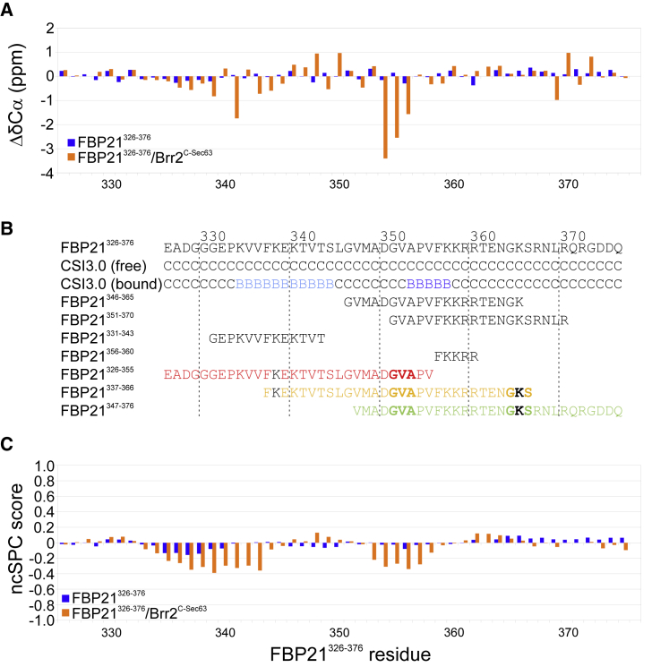
Based on our recent finding that FBP21 regulates human Brr2 helicase activity involved in the activation of the spliceosomal B-complex, we investigated the structural and dynamic contribution of FBP21 to the interaction. By using NMR spectroscopy, we could show that the 50 C-terminal residues of FBP21 (FBP21326-376), which are sufficient to fully form the interaction with the C-terminal Sec63 unit of Brr2 (Brr2C-Sec63), adopt a random-coil conformation in their unbound state. Upon interaction with Brr2C-Sec63, 42 residues of FBP21326-376 cover the large binding site on Brr2C-Sec63 in an extended conformation. Short charged motifs are steering complex formation, still allowing the bound state to retain dynamics. Based on fragment docking in combination with experimental restraints, we present models of the complex structure. The FBP21326-376/Brr2C-Sec63 interaction thus presents an example of an intrinsically disordered protein/ordered-protein interaction in which a large binding site provides high specificity and, in combination with conformational disorder, displays a relatively high affinity.


Figures

Figure 1
Figure 1
Backbone assignment of free and Brr2C-Sec63-bound FBP21326–376. (A) Shown is the assigned 1H-15N-TROSY-HSQC of 140 μM 2H-15N-13C-FBP21326–376 with low signal dispersion, indicating intrinsic disorder. (B) An overlay of 1H-15N-TROSY-HSQC spectrum of 140 μM FBP21326–376 alone and in complex with a threefold molar excess of Brr2C-Sec63. Assigned peaks deriving from FBP21326–376 in the Brr2C-Sec63-bound state are labeled in gray. To see this figure in color, go online.
Figure 2
Figure 2
Secondary structure analysis of free and Brr2C-Sec63-bound FBP21326–376. (A) Cα secondary chemical shifts are shown for free and Brr2C-Sec63-bound FBP21326–376. Fluctuation around zero indicates no defined secondary structure in free FBP21326–376. In the Brr2C-Sec63-bound state, increased negative values indicate regions adopting an extended conformation. (B) The secondary structure was predicted for free and Brr2C-Sec63-bound FBP21326–376 by using the web tool CSI3.0 (14). Free FBP21326–376 is predicted to be disordered (CSI3.0 free), whereas the Brr2C-Sec63-bound state appears to form regions with extended conformation (CSI3.0 bound, light B: interior β-strand, dark B: edge β-strand). Additionally shown are peptide fragments of FBP21326–376 investigated here and/or previously (9). Peptides only used for docking analyses are colored as in Fig. S5, with lysine residues in black indicating the positions of restraints derived from cross-linking and residues in bold indicating the position of restraints derived from NOEs. (C) The secondary structure propensity score (ncSPC score: value 1 corresponds to 100% α-helix, value −1 corresponds to 100% β-strand) was calculated for free and Brr2C-Sec63-bound FBP21326–376 by using the web tool ncSPC (20). To see this figure in color, go online.
Figure 3
Figure 3
Differences in fold and dynamics between free and Brr2C-Sec63-bound FBP21326–376. (A) Plotted are per-residue combined 1H,15N chemical shift differences between the two spectra shown in Fig. 1B. The black line indicates the average chemical shift difference (0.27 ppm). (B) Strips of a 1H-15N-NOESY-HSQC spectrum of 140 μM 2H-15N-13C-FBP21326–376 in the presence of 420 μM Brr2C-Sec63 show HN-HN-NOEs. (C) Heteronuclear 1H-15N-NOE values and T1/T2 relaxation time ratios obtained for 125 μM 15N-FBP21326–376 without or with a threefold molar excess of unlabeled Brr2C-Sec63. Heteronuclear 1H-15N-NOE values represent the average of three independent measurements, error bars reflect the standard deviation. Uncertainties of T1 and T2 times were obtained from exponential fits. To see this figure in color, go online.
Figure 4
Figure 4
Energy-minimized models generated from docking results (Fig. S6C). (A) Models of FBP21326–376 are shown with Brr2C-Sec63 in surface representation. Residues showing chemical shift differences upon addition of FBP21326–376 (9) are shown in strong (if bigger than mean + 1 SD) or light (if between mean and mean + 1 SD) colors. Chemical shift differences are induced by N-terminal residues (that is upon interaction with FBP21326–376 but not with N-terminally truncated fragments) by core residues (that is upon interaction with FBP21326–376, FBP21346–365, and FBP21351–370) or by C-terminal residues (that is upon interaction with FBP21326–376 or FBP21351–370). (BE) PRE experiments confirm the models. (B) and (C) show regions of the overlay of 1H-15N-TROSY-HSQC spectra of 100 μM 15N-Brr2C-Sec63 in complex with equimolar amounts of (B) FBP21S344C-MTSL and (C) FBP21S366C-MTSL in its paramagnetic and its diamagnetic states. (D) and (E) illustrate the position of the label in the models (arrows) of FBP21326–376 and regions experiencing relaxation enhancement in Brr2C-Sec63 in surface representation. Indicated is a loss in peak intensity beyond the average minus SD (67%) or beyond 25% in light or dark colors respectively. (FI) Mutations introduced in FBP21 confirm the models. (F) and (G) show regions of the overlay of 1H-15N-TROSY-HSQC spectra of 100 μM 15N-Brr2C-Sec63 free in complex with a twofold molar excess of FBP21326–376 and a twofold molar excess of (F) FBP21GGG or (G) FBP21ANLA. (H) and (I) show Brr2C-Sec63 for which chemical shift differences upon complex formation with FBP21GGG (H) or FBP21ANLA (I) drops beyond the significance cutoff in the presence of the mutations. The position of the mutations in FBP21326–376 are indicated with an arrow. PDB: 4f91 (23) is used to present Brr2C-Sec63 with models of FBP21326–376. To see this figure in color, go online.

References

    1. Dyson H.J., Wright P.E. Intrinsically unstructured proteins and their functions. Nat. Rev. Mol. Cell Biol. 2005;6:197–208. - PubMed
    1. Tompa P., Fuxreiter M. Fuzzy complexes: polymorphism and structural disorder in protein-protein interactions. Trends Biochem. Sci. 2008;33:2–8. - PubMed
    1. Tompa P. Intrinsically unstructured proteins. Trends Biochem. Sci. 2002;27:527–533. - PubMed
    1. Gunasekaran K., Tsai C.J., Nussinov R. Extended disordered proteins: targeting function with less scaffold. Trends Biochem. Sci. 2003;28:81–85. - PubMed
    1. Korneta I., Bujnicki J.M. Intrinsic disorder in the human spliceosomal proteome. PLoS Comput. Biol. 2012;8:e1002641. - PMC - PubMed

Publication types

MeSH terms

LinkOut - more resources