. 2019 Jan 2;116(1):245-254.
doi: 10.1073/pnas.1817239116. Epub 2018 Dec 17.

Structural determinants driving homoserine lactone ligand selection in the Pseudomonas aeruginosa LasR quorum-sensing receptor

Affiliations

Structural determinants driving homoserine lactone ligand selection in the Pseudomonas aeruginosa LasR quorum-sensing receptor

Amelia R McCready et al. Proc Natl Acad Sci U S A. .

Abstract

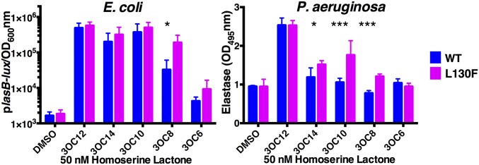
Quorum sensing is a cell-cell communication process that bacteria use to orchestrate group behaviors. Quorum sensing is mediated by signal molecules called autoinducers. Autoinducers are often structurally similar, raising questions concerning how bacteria distinguish among them. Here, we use the Pseudomonas aeruginosa LasR quorum-sensing receptor to explore signal discrimination. The cognate autoinducer, 3OC12 homoserine lactone (3OC12HSL), is a more potent activator of LasR than other homoserine lactones. However, other homoserine lactones can elicit LasR-dependent quorum-sensing responses, showing that LasR displays ligand promiscuity. We identify mutants that alter which homoserine lactones LasR detects. Substitution at residue S129 decreases the LasR response to 3OC12HSL, while enhancing discrimination against noncognate autoinducers. Conversely, the LasR L130F mutation increases the potency of 3OC12HSL and other homoserine lactones. We solve crystal structures of LasR ligand-binding domains complexed with noncognate autoinducers. Comparison with existing structures reveals that ligand selectivity/sensitivity is mediated by a flexible loop near the ligand-binding site. We show that LasR variants with modified ligand preferences exhibit altered quorum-sensing responses to autoinducers in vivo. We suggest that possessing some ligand promiscuity endows LasR with the ability to optimally regulate quorum-sensing traits.

Keywords: LasR; Pseudomonas aeruginosa; crystal structure; homoserine lactone; quorum sensing.


Conflict of interest statement

The authors declare no conflict of interest.

Figures

Fig. 1.
Fig. 1.
LasR is activated by multiple homoserine lactone autoinducers. (A) LasR-dependent bioluminescence was measured in E. coli. Arabinose-inducible LasR was produced from one plasmid and the plasB-lux reporter construct was carried on a second plasmid; 0.1% arabinose was used for LasR induction. (B) Elastase activity was measured from ΔlasI P. aeruginosa using elastin-Congo red as the substrate. In A and B, 100 nM of the designated HSLs were tested. Two technical replicates were performed for each biological sample and three biological replicates were assessed. Error bars denote SDs of the mean. Paired two-tailed t tests were performed comparing each compound to the DMSO control. *P < 0.05, **P < 0.01, ***P < 0.0001. (C) Comparison of LasR LBD protein levels in whole-cell lysates (W) and in the soluble fractions (S) of E. coli cells harboring DNA encoding the LasR LBD on a plasmid; 1 mM IPTG was used for LasR LBD induction and either 1% DMSO or 10 μM of the indicated HSL was supplied. In all lanes, protein from 0.05 OD of cells was loaded. Results are representative of three trials. (D) Thermal-shift analyses of purified LasR LBD bound to 3OC10HSL (Left), 3OC12HSL (Center), and 3OC14HSL (Right) without (designated DMSO) and following supplementation with an additional 10 μM of the indicated HSLs. Normalized fluorescence represents the first derivative of the raw fluorescence data (59). Each line represents the average of three replicates.
Fig. 2.
Fig. 2.
LasR S129 mutants alter LasR responses to HSL autoinducers. (A) Wild-type LasR and LasR S129 mutant bioluminescence from the E. coli plasB-lux reporter; 1 μM of the indicated HSLs were provided (see Fig. 1A for detail). (B) Wild-type LasR and LasR S129F-driven elastase activity in ΔlasI P. aeruginosa; 10 μM of the indicated HSLs were provided (see Fig. 1B for detail). In A and B, two technical replicates were performed for each biological sample and three biological replicates were assessed. Error bars denote SDs of the mean. Unpaired t tests were performed to compare each mutant LasR-HSL combination to that of wild-type LasR with the same compound. Due to space constraints, statistics for A are provided in SI Appendix, Table S3. **P < 0.01, ***P < 0.0001. (C) Comparison of LasR S129F LBD protein levels in whole-cell lysates (W) and in the soluble fractions (S) of E. coli cells harboring DNA encoding the LasR S129F LBD on a plasmid; 1 mM IPTG was used for LasR S129F LBD induction and either 1% DMSO or 10 μM of the indicated HSL was supplied. In all lanes, protein from 0.05 OD of cells was loaded. Results are representative of three trials. (D) Thermal-shift analyses of purified LasR S129F LBD bound to 3OC12HSL (Left) and 3OC14HSL (Right) without (designated DMSO) and following supplementation with an additional 10 μM of the indicated HSLs. Normalized fluorescence represents the first derivative of the raw fluorescence data (59). Each line represents the average of three replicates.
Fig. 3.
Fig. 3.
LasR L130F displays enhanced responses to HSL autoinducers. (Left) Wild-type LasR and LasR L130F-driven bioluminescence from the E. coli plasB-lux reporter; 50 nM of the indicated HSLs were provided (see Fig. 1A for detail). (Right) Wild-type LasR and LasR L130F-driven elastase activity in ΔlasI P. aeruginosa; 50 nM of the indicated HSLs were provided (see Fig. 1B for detail). In both A and B, two technical replicates were performed for each biological sample and three biological replicates were assessed. Error bars denote SDs of the mean. Unpaired t tests were performed to compare each LasR L130F-HSL combination to that of wild-type LasR with the same compound. *P < 0.05, ***P < 0.0001.
Fig. 4.
Fig. 4.
The LasR LBD L130F is more stable than the wild-type LasR LBD. (A) Thermal-shift analyses of purified LasR LBD (solid lines) and LasR LBD L130F (dotted lines) bound to 3OC12HSL (orange) or 3OC14HSL (green). Each line represents the average of three replicates. (B) Thermal-shift analyses of purified LasR LBD L130F bound to 3OC8HSL (Upper Left), 3OC10HSL (Lower Left), 3OC12HSL (Upper Right), and 3OC14HSL (Lower Right) without (designated DMSO) and following supplementation with an additional 10 μM of the indicated HSLs. In A and B, normalized fluorescence represents the first derivative of the raw fluorescence data (59). (C) Comparison of LasR L130F levels in the whole-cell lysates (W) and the soluble fractions (S) of E. coli cells harboring DNA encoding LasR LBD L130F on a plasmid (see Fig. 1C for details). Either 1% DMSO or 10 μM of the indicated HSL molecule was added.
Fig. 5.
Fig. 5.
Wild-type LasR, LasR S129F, and LasR L130F display distinct pyocyanin production phenotypes in response to high and low concentrations of 3OC12HSL. Pyocyanin production was measured in ΔlasI P. aeruginosa strains over the growth curve. The y axis “Pyocyanin” is the amount of pyocyanin pigment (OD695 nm) over cell density (OD600 nm). Designations are: red, DMSO control; blue, wild-type LasR; green, LasR S129F; pink, LasR L130F. Concentrations used are: (Left) 50 nM 3OC12HSL, (Right) 1 μM 3OC12HSL. Data show the mean of three biological replicates. Error bars denote SDs of the mean. Two-way ANOVAs were performed to compare LasR S129F and LasR L130F to wild-type LasR under each condition. A two-way ANOVA was also performed to compare wild-type LasR with 3OC12HSL and with DMSO alone. *P < 0.05, ***P < 0.0001.
Fig. 6.
Fig. 6.
Crystal structures of LasR LBD L130F bound to 3OC10HSL and 3OC14HSL. (A) Crystal structures of LasR LBD L130F:3OC10HSL (gold) and LasR LBD L130F:3OC14HSL (magenta) compared with the wild-type LasR LBD:3OC12HSL structure [blue, modified from PDB: 2UV0 (29)]. The Lower images show 90° rotations of the crystal structures relative to the images above. In the two Upper rightmost structures, the asterisks highlight the LasR loop region that includes residues 40–51 and that undergoes a conformational shift when 3OC14HSL is bound. (B) Structural comparison of LasR LBD:3OC12HSL (protein: blue, ligand: orange; modified from PDB ID code 2UVO) (29) and LasR LBD L130F:3OC10HSL crystal structures (protein: gold, ligand: magenta). Amino acids drawn in stick format show important residues for lactone head binding (Y56, W60, R61, D73, T75, W88, Y93, and S129) and acyl chain binding (G38, L40, A50, I52, A70, V76, L125, and A127). (C) Structural comparison of LasR LBD:3OC12HSL (protein: blue, ligand: orange; modified from PDB ID code 2UVO) (29) and LasR LBD L130F:3OC14HSL crystal structures (protein: magenta, ligand: green). Amino acids drawn in stick format are the same as in B. Note the shift in the position of the loop region corresponding to residues 40–51.
Fig. 7.
Fig. 7.
A conserved flexible loop region confers promiscuity to LuxR-type receptors. (A) Upper images: structural comparison of the LuxR-type receptors LasR LBD L130F:3OC14HSL (magenta) and SdiA LBD:3OC8HSL [green, modified from PDB: 4Y17 (21) PDB ID code AY17] that exhibit promiscuity with respect to ligand binding. Lower images: structural comparison of the LuxR-type receptors CviR LBD:C6HSL [pink, modified from PDB: 3QP1 (39)] and TraR LBD:3OC8HSL [silver, modified from PDB: 1L3L (40)] that display strict ligand specificity. (B) Structural comparison of the protein:ligand interfaces for LasR LBD L130F:3OC14HSL (Upper Left, magenta), SdiA LBD:3OC8HSL (Upper Right, green), CviR LBD:C6HSL (Lower Left, pink), and TraR LBD:3OC8HSL (Lower Right, silver). Residues that make important hydrophobic side chain interactions in each protein:ligand complex are shown in stick format and named.
Fig. 8.
Fig. 8.
LasR A127W occludes long chain HSLs from the binding pocket. E. coli plasB-lux reporter assays were performed with wild-type LasR and LasR A127W in response to 3OC12HSL, 3OC10HSL, 3OC8HSL, and 3OC6HSL. EC50 values were obtained by performing dose–response assays for each HSL and analyzing the resulting data using nonlinear regression analysis. Two technical replicates were performed for each biological sample and at least three biological replicates were assessed. In the case of wild-type LasR, three biological replicates were performed for each experiment and then combined across all assays.

Similar articles

Cited by

References

    1. Papenfort K, Bassler BL. Quorum sensing signal-response systems in Gram-negative bacteria. Nat Rev Microbiol. 2016;14:576–588. - PMC - PubMed
    1. Albus AM, Pesci EC, Runyen-Janecky LJ, West SE, Iglewski BH. Vfr controls quorum sensing in Pseudomonas aeruginosa. J Bacteriol. 1997;179:3928–3935. - PMC - PubMed
    1. Latifi A, et al. Multiple homologues of LuxR and LuxI control expression of virulence determinants and secondary metabolites through quorum sensing in Pseudomonas aeruginosa PAO1. Mol Microbiol. 1995;17:333–343. - PubMed
    1. Engebrecht J, Nealson K, Silverman M. Bacterial bioluminescence: Isolation and genetic analysis of functions from Vibrio fischeri. Cell. 1983;32:773–781. - PubMed
    1. Hanzelka BL, et al. Acylhomoserine lactone synthase activity of the Vibrio fischeri AinS protein. J Bacteriol. 1999;181:5766–5770. - PMC - PubMed

Publication types

MeSH terms

LinkOut - more resources