. 2019 Jan 2;146(1):dev169219.
doi: 10.1242/dev.169219.

Targeted substrate degradation by Kelch controls the actin cytoskeleton during ring canal expansion

Affiliations

Targeted substrate degradation by Kelch controls the actin cytoskeleton during ring canal expansion

Andrew M Hudson et al. Development. .

Abstract

During Drosophila oogenesis, specialized actin-based structures called ring canals form and expand to accommodate growth of the oocyte. Previous work demonstrated that Kelch and Cullin 3 function together in a Cullin 3-RING ubiquitin ligase complex (CRL3Kelch) to organize the ring canal cytoskeleton, presumably by targeting a substrate for proteolysis. Here, we use tandem affinity purification followed by mass spectrometry to identify HtsRC as the CRL3Kelch ring canal substrate. CRISPR-mediated mutagenesis of HtsRC revealed its requirement in the recruitment of the ring canal F-actin cytoskeleton. We present genetic evidence consistent with HtsRC being the CRL3Kelch substrate, as well as biochemical evidence indicating that HtsRC is ubiquitylated and degraded by the proteasome. Finally, we identify a short sequence motif in HtsRC that is necessary for Kelch binding. These findings uncover an unusual mechanism during development wherein a specialized cytoskeletal structure is regulated and remodeled by the ubiquitin-proteasome system.

Keywords: Actin cytoskeleton; Cullin 3; Drosophila oogenesis; Kelch; Ring canal; Ubiquitin-proteasome system.


Conflict of interest statement

Competing interestsThe authors declare no competing or financial interests.

Figures
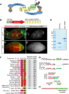
Fig. 1.
Fig. 1.
Tandem affinity purification and mass spectrometry reveals HtsRC as a potential CRL3Kelch substrate. (A) Cartoon of a CRL3Kelch dimer, showing ubiquitylation of a substrate (green circle). (B) Diagram of TAP-tagged mCherry fused to the Kelch KREP domain. (C,C′) High-level expression of the mCherry::KREP fusion using the matGal4 driver resulted in ring canal localization and a dominant kelch-like phenotype. (D,D′) In cher1 mutant ovaries, the mCherry::KREP fusion exhibited a homogeneous cytosolic distribution and was not associated with ring canals. (E) Proteins bound to TAP-tagged mCherry or TAPmCherry::KREP were purified from cher1 mutant ovaries, separated on a 4-12% SDS-PAGE gel, stained with colloidal Coomassie and subjected to LC-MS/MS. Purified TAPmCherry and TAPmCherry::KREP proteins are indicated by red arrowheads. (F) Sequence analysis of proteins bound specifically to TAPmCherry::KREP that contain a motif consisting of several acidic residues followed by a PEAEQ consensus sequence defined by the expression [PLT]-E-[AD]-[DE]-[QD]. Sequences were manually aligned in JalView (Waterhouse et al., 2009). (G) Alternative splicing of hts transcripts produces several mRNAs, including mRNAs encoding Adducin. The ovary-specific ovhts mRNA contains exon 12 (green) and produces a polyprotein that is cleaved to produce truncated Adducin (HtsF) that localizes to fusomes and the novel HtsRC protein that localizes to ring canals. (H) Peptides identified from proteins bound to mCherry::KREP mapped entirely to HtsRC (yellow boxes). See also Figs S1, S2 and Table S1.
Fig. 2.
Fig. 2.
HtsRC is ubiquitylated and degraded by the proteasome. (A) Cartoon showing full-length 3xHA::HtsRC::HA and cleaved HtsRC::HA protein products. (B) S2 cells expressing 3xHA::HtsRC::HA were treated with DMSO (control) or 1 µM bortezomib for 3 or 6 h, 24 h post-transfection, lysed and analyzed by western blotting. Proteasome inhibition results in higher molecular weight, presumably ubiquitylated, HtsRC species. (C) S2 cells were treated with DMSO (control) or 1 µM bortezomib for 3 and 6 h, 36 h post-transfection, lysed and analyzed by western blotting. (D) Levels of HtsRC::HA were elevated after bortezomib treatment. *P<0.05; Student's t-test. Data are from three independent experiments. (E) Cells expressing 3xHA::HtsRC::HA 24 h post-transfection were treated with 100 µg/ml cycloheximide (CHX) with or without 1 µM bortezomib, harvested at 3 and 6 h timepoints, and analyzed by western blotting. (F) Quantification of HtsRC protein levels from three independent CHX chase experiments. HtsRC protein levels decreased after CHX treatment and were stabilized with bortezomib treatment, suggesting that HtsRC is degraded by the proteasome. *P<0.05; Student's t-test. (G) Blots of proteins eluted from Ni++-NTA purifications from control ovary extracts lacking His::Ub, and wild-type or kelchDE1 mutant extracts expressing His::Ub. Anti-ubiquitin antibody P4D1 revealed strong and specific enrichment of ubiquitylated proteins in purifications from His::Ub extracts. (H) Blot of total ovarian protein (lane 1) and proteins eluted from Ni++-NTA purifications as in G. Anti-HtsRC antibody revealed a ladder of ubiquitylated HtsRC species in His::Ub purifications from both wild-type and kelch mutant extracts. Two minor cross-reacting bands eluted from Ni++-NTA beads in wild-type extracts were recognized by HtsRC in the control.
Fig. 3.
Fig. 3.
Altered HtsRC expression can suppress or enhance the kelch-like ring canal phenotype. (A,B) Ring canal F-actin thickness in stage 6 (A) or stage 9 (B) egg chambers was calculated using the full width at half max (FWHM) measure of F-actin intensity plots spanning across ring canals. Colored boxed images show representative ring canals of each genotype, with corresponding individual data points below for each ring canal measured. Bars represent the F-actin thickness as mean±95% confidence interval. n denotes number of ring canals measured. (A) Increased or decreased HtsRC expression enhanced or suppressed, respectively, the kelch-like ring canal phenotype observed upon germline-specific proteasome inhibition by RNAi. Proteasome inhibition led to kelch-like ring canals, marked by significantly thicker F-actin rings (red). Increased HtsRC::GFP expression (green) and increased HtsRC::GFP expression with loss of one copy of kelch (yellow) significantly increased ring canal F-actin thickness, whereas removal of one copy of htsRC fully suppressed the kelch-like phenotype observed upon proteasome inhibition (blue). Scale bar: 1 µm. (B) Germline-specific knockdown of Cul3 by RNAi led to kelch-like ring canals (red), marked by significantly thicker F-actin rings. Removal of one copy of htsRC was sufficient to suppress the kelch-like phenotype (blue). ****P<0.0001, ***P<0.001, *P<0.05; one-way ANOVA, Tukey's multiple comparison test. n.s., not significant. Scale bar: 2 µm.
Fig. 4.
Fig. 4.
Genetic analysis indicates hts is epistatic to kelch. (A) Diagram summarizing the locations of CRISPR/NHEJ-induced deletion mutations in hts exon 12. htsI775fs is a 2 bp deletion (Chr2R:19401008…19401009, FlyBase release 6); translation of the mutant cDNA would produce a 62 amino acid extension in the new reading frame starting at I775 of Ovhts. htsT776fs is a single base pair deletion (Chr2R:19401007) resulting in a frameshift extension of 28 amino acids beginning at T776. htsR913fs is a 559 bp deletion (Chr2R:19400036…19400594) resulting in a frameshift extension of 10 amino acids beginning at R913, immediately prior to the DRERPEAEQ sequence. (B,D,F,H) Wild-type, kelDE1, htsR913fs and htsR913fs, kelDE1 stage 10 egg chambers showing the distributions of F-actin, phosphotyrosine (PY) and Filamin (FLN). kelch mutant egg chambers displayed a fully penetrant ‘small oocyte’ phenotype at stage 10 (D). Oocytes in htsR913fs (F) and htsR913fs, kelDE1 double mutant egg chambers (H) did not exhibit a penetrant stage 10 growth defect. (C-C‴,E-E‴,G-G‴,I-I‴) High-resolution images of ring canals from the genotypes indicated. kelDE1and htsR913fs ring canals exhibited opposite phenotypes with respect to ring canal F-actin accumulation: excess F-actin in kelDE1 and only a small amount of peripheral F-actin accumulation around the ring canals in the htsR913fs mutant. Scale bars: 50 µm in H; 5 µm in I‴. (J) Quantification of egg lengths from the indicated genotypes. The htsR913fs, kelDE1 double mutant phenotype was indistinguishable from htsR913fs, demonstrating that hts is epistatic to kelch. Bars represent egg length as mean±95% confidence interval. One-way ANOVA, Tukey's multiple comparison test; n.s., not significant. See also Fig. S4.
Fig. 5.
Fig. 5.
HtsRC ring canal protein levels are dependent on Kelch. (A) Wild-type HtsRC expression in the germarium and subsequent stages. (B) matGal4-driven expression of a shRNA against hts resulted in near elimination of HtsRC from the germline in stages where matGal4 expression was high (yellow dotted line indicates matGal4 expression pattern). (C) Mutations in kelch suppressed the hts shRNA knockdown phenotype, resulting in detectable HtsRC in stage 2-8 egg chambers. The image is a mosaic created from two images using the MosaicJ plug-in from FIJI. (D) HtsRC::GFP fluorescent protein levels decreased with mCherry::Kelch co-expression compared with mCherry control expression. All constructs were driven by matGal4. Individual egg chambers are outlined in yellow. (E) Quantification of HtsRC::GFP fluorescent protein levels at ring canals. ****P<0.0001; Student's t-test. (F) HtsRC::Venus fluorescence levels at ring canals increased in a dose-dependent manner depending on kelch gene copy. Individual egg chambers are outlined in yellow. (G) Quantification of HtsRC::Venus fluorescent protein levels at ring canals. The mean maximum fluorescence intensity of the ovhts::Venus x2 sample (black) was normalized to 100 relative fluorescence units. For E and G, data points in the scatter plot represent the maximum fluorescence intensity of HtsRC::Venus or HtsRC::GFP for each ring canal measured. Bars represent the maximum fluorescence intensity as mean±95% confidence interval of HtsRC::Venus or HtsRC::GFP. n denotes number of ring canals analyzed. ****P<0.0001; one-way ANOVA, Tukey's multiple comparison test. See also Fig. S5.
Fig. 6.
Fig. 6.
Removal of the Kelch NTR results in hyper-active Kelch. (A) Cartoon of Kelch protein motifs, with low-complexity regions (LC) in the NTR (pink boxes). (B,B′) Control flies expressing mCherry under control of the matGal4 driver revealed the expression domain of matGal4 (blue) and had the wild-type pattern of HtsRC accumulation in young egg chambers. (C,C′) Expression of wild-type Kelch protein with the matGal4 driver resulted in a normal distribution of HtsRC in egg chambers. (D,D′) matGal4-driven expression of KelchΔNTR resulted in dramatic loss of HtsRC fluorescence compared with expression of wild-type Kelch. Yellow dotted line indicates the expression domain of matGal4; HtsRC levels in wild type and matGal4>kelchΔNTR were comparable in the germarium, where the matGal4 driver is not expressed. (E) Representative western blots showing levels of HtsRC and Kelch. A Tubulin immunoblot is shown as a loading control. (F) Expression of KelchΔNTR driven by otuGal4 resulted in reduced HtsRC protein compared with w1118 or matGal4>kelchWT, despite higher levels of Kelch in matGal4>kelchWT (see G). *P<0.05. (G) KelchΔNTR driven by otuGal4 resulted in approximately twice the level of endogenous Kelch. matGal4-driven expression of wild-type Kelch resulted in an approximately fivefold increase in Kelch expression.
Fig. 7.
Fig. 7.
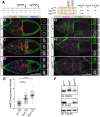
A PEAEQ sequence motif in HtsRC is necessary for HtsRC interaction with Kelch. (A) Location of sgRNAs targeting the HtsRC PEAEQ motif (left) and summary of single-codon deletion mutations in PEAEQ sequence (middle). Red arrowheads indicate locations of sgRNA-directed Cas9-mediated cleavages. htsΔE992 is an in-frame deletion resulting in a single amino acid deletion of the conserved E922 residue; htsΔQ925 deletes Q925. Fertility and relative HtsRC and Kelch immunofluorescence levels at ring canals are summarized on the right. (B-D) Control (w1118), htsΔE992 and kelDE1 egg chambers labeled to reveal F-actin, HtsRC and Filamin. Homozygous htsΔE992 egg chambers and ring canals were indistinguishable from kelDE1. (E) Measured fluorescence intensity of HtsRC immunolabeled ring canals. Bars represent the fluorescence intensity as mean±95% confidence interval. ****P<0.0001; one-way ANOVA with Dunnett's multiple comparison test. (F-I) Kelch localization in control (w1118), htsΔE992 and kelDE1 egg chambers. Kelch localized to ring canals in wild-type egg chambers (G), but not in htsΔE992 mutants (I), similar to kelch loss-of-function mutations (H), despite being expressed at normal levels (F). For B-D and G-I, ring anals within yellow box are shown in the inset to the right. Scale bar: 5 µm in inset. (F) Western blot showing steady-state levels of HtsRC and Kelch; Tubulin serves as a loading control. Overall HtsRC levels were not significantly increased compared with wild type. See also Fig. S6.
Fig. 8.
Fig. 8.
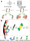
Model for ring canal growth regulated by CRL3Kelch. (A) Wild-type (WT) and kelch ring canals diagrammed in cross-section. Ring canals recruit HtsRC, which results in the accumulation of a robust F-actin cytoskeleton that supports ring canal growth. (B) In wild type, CRL3Kelch ubiquitylates HtsRC, targeting it for destruction by the proteasome. Removal of HtsRC by CRL3Kelch-mediated destruction is required for the disassembly of the ring canal cytoskeleton from the ring canal lumen as the ring canal expands.

Similar articles

Cited by

References

    1. Adams J., Kelso R. and Cooley L. (2000). The kelch repeat superfamily of proteins: propellers of cell function. Trends Cell Biol. 10, 17-24. 10.1016/S0962-8924(99)01673-6 - DOI - PubMed
    1. Arama E., Bader M., Rieckhof G. E. and Steller H. (2007). A ubiquitin ligase complex regulates caspase activation during sperm differentiation in Drosophila. PLoS Biol. 5, e251 10.1371/journal.pbio.0050251 - DOI - PMC - PubMed
    1. Bastock R. and St Johnston D. (2008). Drosophila oogenesis. Curr. Biol. 18, R1082-7 10.1016/j.cub.2008.09.011 - DOI - PubMed
    1. Bomont P., Cavalier L., Blondeau F., Ben Hamida C., Belal S., Tazir M., Demir E., Topaloglu H., Korinthenberg R., Tüysüz B. et al. (2000). The gene encoding gigaxonin, a new member of the cytoskeletal BTB/kelch repeat family, is mutated in giant axonal neuropathy. Nat. Genet. 26, 370-374. 10.1038/81701 - DOI - PubMed
    1. Boyden L. M., Choi M., Choate K. A., Nelson-Williams C. J., Farhi A., Toka H. R., Tikhonova I. R., Bjornson R., Mane S. M., Colussi G. et al. (2012). Mutations in kelch-like 3 and cullin 3 cause hypertension and electrolyte abnormalities. Nature 482, 98-102. 10.1038/nature10814 - DOI - PMC - PubMed

Publication types

LinkOut - more resources