. 2019 Feb 15;294(7):2318-2328.
doi: 10.1074/jbc.RA118.006106. Epub 2018 Dec 17.

Retinal degeneration 3 (RD3) protein, a retinal guanylyl cyclase regulator, forms a monomeric and elongated four-helix bundle

Affiliations

Retinal degeneration 3 (RD3) protein, a retinal guanylyl cyclase regulator, forms a monomeric and elongated four-helix bundle

Igor V Peshenko et al. J Biol Chem. .

Abstract

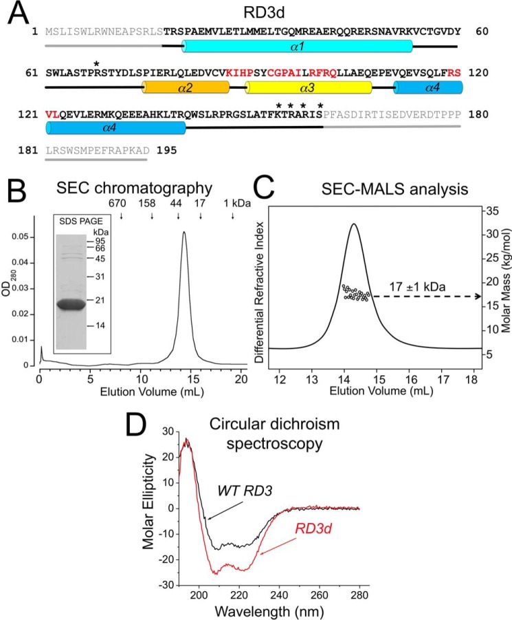
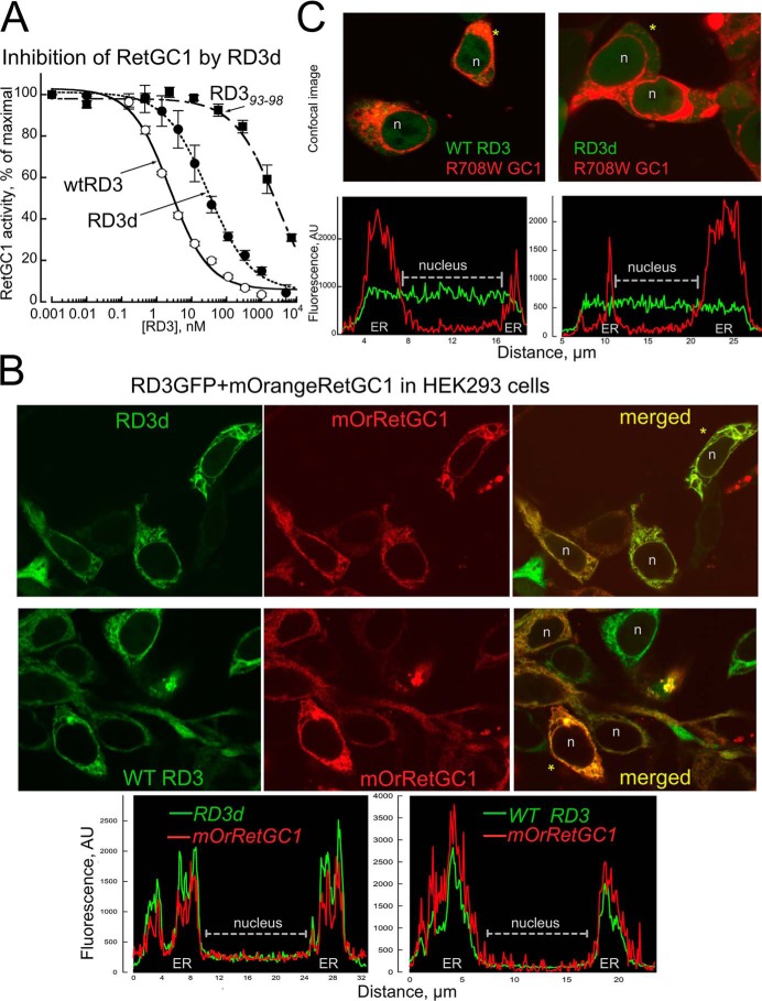
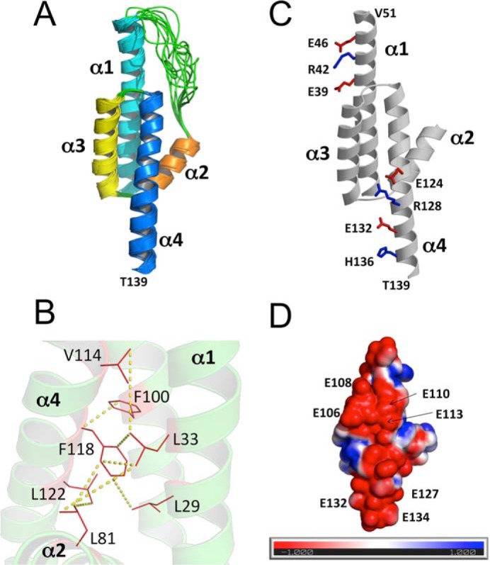
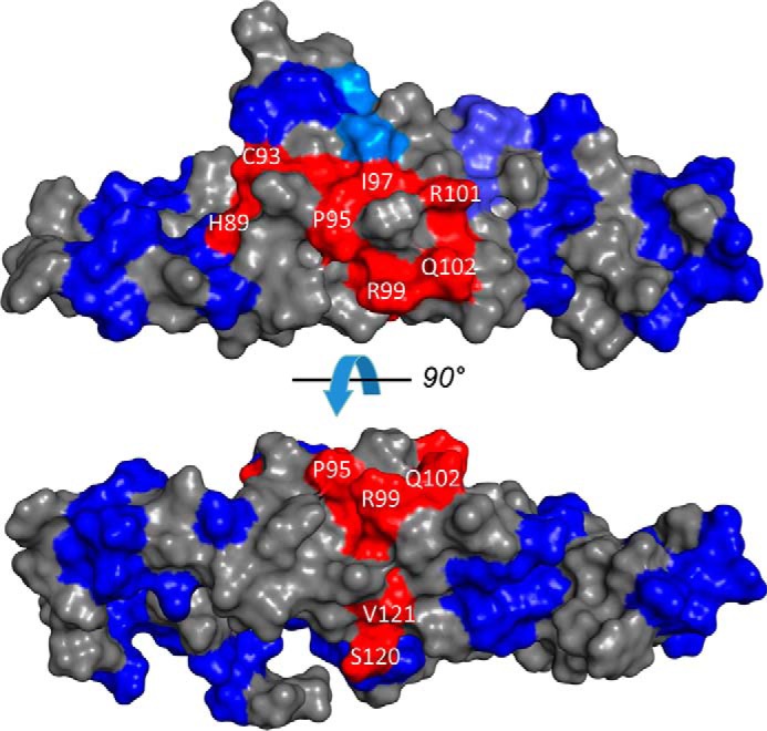
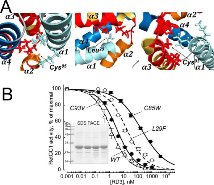
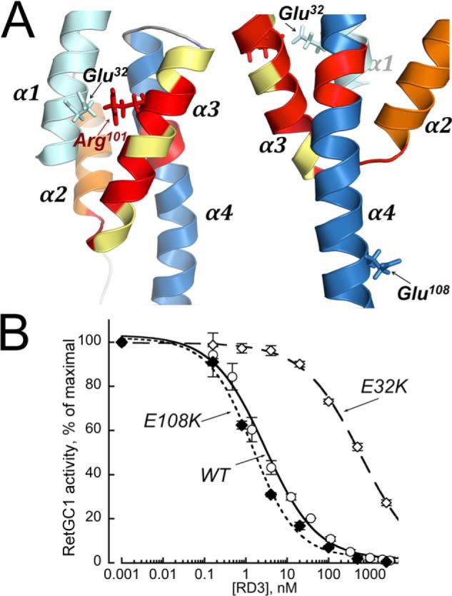
Retinal degeneration 3 (RD3) protein promotes accumulation of retinal membrane guanylyl cyclase (RetGC) in the photoreceptor outer segment and suppresses RetGC activation by guanylyl cyclase-activating proteins (GCAPs). Mutations truncating RD3 cause severe congenital blindness by preventing the inhibitory binding of RD3 to the cyclase. The high propensity of RD3 to aggregate in solution has prevented structural analysis. Here, we produced a highly soluble variant of human RD3 (residues 18-160) that is monomeric and can still bind and negatively regulate RetGC. The NMR solution structure of RD3 revealed an elongated backbone structure (70 Å long and 30 Å wide) consisting of a four-helix bundle with a long unstructured loop between helices 1 and 2. The structure reveals that RD3 residues previously implicated in the RetGC binding map to a localized and contiguous area on the structure, involving a loop between helices 2 and 3 and adjacent parts of helices 3 and 4. The NMR structure of RD3 was validated by mutagenesis. Introducing Trp85 or Phe29 to replace Cys or Leu, respectively, disrupts packing in the hydrophobic core and lowers RD3's apparent affinity for RetGC1. Introducing a positive charge at the interface (Glu32 to Lys) also lowered the affinity. Conversely, introducing Val in place of Cys93 stabilized the hydrophobic core and increased the RD3 affinity for the cyclase. The NMR structure of RD3 presented here provides a structural basis for elucidating RD3-RetGC interactions relevant for normal vision or blindness.

Keywords: guanylate cyclase (guanylyl cyclase); nuclear magnetic resonance (NMR); photoreceptor; phototransduction; retinal degeneration.


Conflict of interest statement

The authors declare that they have no conflicts of interest with the contents of this article

Figures

Figure 1.
Figure 1.
A, primary structure of the hRD3. The residues that were deleted from the soluble RD3d form are shown in gray; residues 18–160 are highlighted in bold; the asterisks mark residues replaced by negatively charged side chains; α1–α4 cylinders mark α-helical structures; and straight lines mark the unstructured regions, either predicted (gray) or found in the NMR structure (black). Residues in the regions previously mapped as parts of the cyclase-binding interface in RD3 (14) are highlighted in red. B, size-exclusion chromatography of the purified RD3d suggests dimerization or nonspherical shape of the protein. RD3 purified from E. coli as described under “Experimental procedures” was chromatographed on a Superdex 200 HR column. The vertical arrows mark the peak elution volumes for molecular mass standards (Bio-Rad chromatography standards: thyroglobulin, IgG, ovalbumin, myoglobin, and vitamin B12). Note that the 18-kDa RD3d elutes in a volume corresponding to a 32-kDa globular protein. Inset, 15% SDS-PAGE of the purified RD3d, Coomassie Blue R-250 stain. C, SEC-MALS analysis of RD3. The molar mass of RD3 in solution (circles) was calculated from a Zimm plot analysis of the observed light scattering intensity using a refractive index increment, dn/dc = 0.185 liter g−1 (16, 50). The protein concentration was 200 μm. D, CD spectra of RD3d (red trace) and full-length WT RD3 (black trace). Molar ellipticity is plotted along the y axis in units of mdeg cm2 dmol−1. A quantitative spectral analysis indicates that RD3d secondary structure is comprised of 65% α-helix and 35% random coil compared with 55% α-helix and 45% random coil for WT RD3. The larger percentage of random coil for WT RD3 is consistent with the truncated regions (residues 1–17 and 161–195) adopting an unstructured random coil.
Figure 2.
Figure 2.
RD3d retains the ability to regulate RetGC1. A, the RD3d expressed in E. coli inhibits RetGC1–GCAP1 complex in submicromolar range in vitro. The activity of a recombinant RetGC1 reconstituted with 1.5 μm purified GCAP1 was assayed at different concentrations of WT RD3 (●), RD3d (○), and full-size RD3 with the scrambled residues 93–98 (14) (■) and normalized per maximal activity of the cyclase assayed in the absence of RD3; data (mean ± S.D., n = 3) are fitted using a Synergy Kaleidagraph software assuming a sigmoidal function, A% = 100/(1 + ([RD3]/EC50)h), where h is a Hill coefficient. For WT, RD3d, and RD393–98, the EC50 values were 3, 36, and 2386 nm, and the Hill coefficients were 0.78 ± 0.09, 0.71 ± 0.07, and 0.69 ± 0.2, respectively. B and C, RD3d-GFP co-localiziation with mOrange RetGC1 in HEK293 cells. B, confocal images of RD3d-GFP (top) and WT RD3-GFP (middle) co-expressed with mOrange RetGC1 in HEK293 cells, and fluorescence distribution profiles for both tags (bottom) across the cells marked with asterisks. Note that both RD3 and RetGC1 in each case co-localize predominantly in the endoplasmic reticulum (ER) membranes and are void from the nucleus (marked n in B and C). C, neither RD3d nor WT RD3 bind W708R RetGC1 mutant. Confocal images (top) of WT RD3-GFP (left) and RD3d-GFP (right) co-expressed with W708R mOrange RetGC1 and the respective fluorescence profiles (bottom). Note that both forms of RD3-GFP are uniformly distributed throughout the cytoplasm and the nucleus of the cells and does not co-localize with the mutant cyclase.
Figure 3.
Figure 3.
NMR-derived structures of RD3d. A, ensemble of 10 lowest energy NMR structures of RD3d (Protein Data Bank code 6DRF). Main-chain structures are depicted by a ribbon diagram. Structural statistics are given in Table 1. B, energy-minimized average structure of RD3d, showing the side-chain atoms of residues in the hydrophobic core. Yellow dashed lines show representative long-range NOE distances measured between residues in the hydrophobic core. C, RD3d salt-bridge interactions in helices α1 and α4 rigidify the elongated RD3 structure. Positively and negatively charged side-chain atoms are depicted by sticks and are colored blue and red, respectively. D, surface representation of RD3d showing the electrostatic potential of solvent-accessible surface residues with nearly the same view as in C. Negatively charged surface is highlighted red, and positively charged surface is blue. Exposed glutamate residues are indicated that form an extended negatively charged patch.
Figure 4.
Figure 4.
The RetGC interface localizes to the central part of the RD3 surface with a similar view as in Fig. 3C rotated 90° counterclockwise. Fragments of RD3 primary structure implicated in the inhibitory binding of RD3 to RetGC1 (red) and those that sustain mutagenesis without loss of the inhibitory binding to the cyclase (blue) (14) are superimposed on the RD3 NMR model.
Figure 5.
Figure 5.
A, residues Cys85 (left), Leu29 (middle), and Cys93 (right) were replaced in a full-length RD3 with the respective Trp, Phe, or Val to modify the interactions between residues proximal to the RetGC-binding domain in the NMR structure of the RD3d (marked in red). The C85W and L29F, inserting overly large residues, are expected to be unfavorable for the packing of the cyclase-binding domain, whereas the C93V is expected to stabilize the interactions within the domain. B, the activity of a recombinant RetGC1 reconstituted with 1.5 μm purified GCAP1 was assayed at different concentrations of the WT (●), C85W (■), L29F (○), and C93V (▵) full-length RD3 and normalized per maximal activity of the cyclase assayed in the absence of RD3; data were normalized and fitted as described in Fig. 2A. The EC50 values for the WT, C85W, L29F, and C93V RD3 from the fit were 3.4, 184, 17.3, and 1.3 nm, with a negative (0.5 < h ≤ 0.7) cooperativity in all cases. Inset, 15% SDS-PAGE of the RD3 variants used in the experiment. Lanes left to right show molecular mass standards, WT RD3, C85W, C93V, and L29F; Coomassie Blue stain.
Figure 6.
Figure 6.
Differential effects of replacing charges in two surface exposed residues in a full-length RD3. A, Glu32 in helix α1 (left), proximal to Arg101 located in the putative RetGC-binding interface part (14) of α3, or Glu108 in helix α4, facing away from the putative cyclase-binding interface (right), was replaced by positively charged Lys. B, the activity of a recombinant RetGC1 reconstituted with 1.5 μm purified GCAP1 was assayed at different concentrations of the full-length WT (○), E108K (♦), or E32K (♢) RD3 as described in Fig. 2A. The respective EC50 values for the WT, E108K, and E32K were 2.8, 1.5, and 594 nm with a negative (0.6 < h < 0.8) cooperativity in all cases.

References

    1. Arshavsky V. Y., and Burns M. E. (2014) Current understanding of signal amplification in phototransduction. Cell. Logist. 4, e29390 10.4161/cl.29390 - DOI - PMC - PubMed
    1. Arshavsky V. Y., Lamb T. D., and Pugh E. N. (2002) G proteins and phototransduction. Annu. Rev. Physiol. 64, 153–187 10.1146/annurev.physiol.64.082701.102229 - DOI - PubMed
    1. Koch K. W., and Dell'Orco D. (2015) Protein and signaling networks in vertebrate photoreceptor cells. Front. Mol. Neurosci. 8, 67 - PMC - PubMed
    1. Dizhoor A. M., Olshevskaya E. V., and Peshenko I. V. (2010) Mg2+/Ca2+ cation binding cycle of guanylyl cyclase activating proteins (GCAPs): role in regulation of photoreceptor guanylyl cyclase. Mol. Cell Biochem. 334, 117–124 10.1007/s11010-009-0328-6 - DOI - PMC - PubMed
    1. Dizhoor A. M., Lowe D. G., Olshevskaya E. V., Laura R. P., and Hurley J. B. (1994) The human photoreceptor membrane guanylyl cyclase, RetGC, is present in outer segments and is regulated by calcium and a soluble activator. Neuron 12, 1345–1352 10.1016/0896-6273(94)90449-9 - DOI - PubMed

Publication types

MeSH terms

Associated data